Monthly Archives: January 2013

Earthling Bulletin #13

by Piter Kehoma Boll
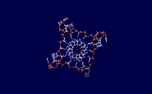
For the first time, the strange four-strand DNA has been found in cells! Picture by Jean-Paul Rodriguez, extracted from nature.com

For the first time, the strange four-strand DNA has been found in cells! Picture by Jean-Paul Rodriguez, extracted from nature.com

News

Blog

Art & Books

Scientific Articles

Leave a comment

Filed under Bulletins

Where did milk come from? The mysterious origin of lactation in mammals.

by Piter Kehoma Boll

ResearchBlogging.org In previous posts, I talked about two scientists who introduced “revolutionary” ideas to explain certain aspects in evolution, contradicting what other specialists use to say. But they come up with such unlikely explanations and use to ignore anything that says something contrary to their ideas that no one takes them seriously. You can read about them here and here.

Today, however, I will talk about another scientist who had interesting and innovative ideas, but much more likely and based on many more evidences. His name is Olav T. Oftedal and the topic is the origin of lactation.

As everybody knows, mammals are characterized mainly by the fact that females nourish their young through mammary secretions, the thing we know as “milk”. But how did this trace evolved in the mammalian lineage?

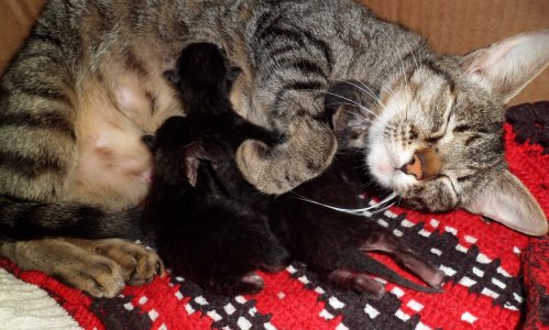
A cat (Nani) with her babies. Photo by Piter Kehoma Boll

A cat (Nani) with her babies. Photo by Piter Kehoma Boll*

Charles Darwin was one of the first ones to think of an explanation. Based on marsupials, Darwin concluded that mammals evolved from animals which carried eggs in a pouch and nourished them by cutaneous secretions. His idea was also inspired by seahorses, were eggs are carried in an abdominal pouch by the male and it was though that they might be nourished by cutaneous secretions too. However the subsequent discovery that the platypus, one the most primitive mammals, lays shelled eggs and have no pouch marked the ruin of Darwin’s theory on the origin of lactation. But… did it indeed?

In a paper published in 2002 in the Journal of Mammary Gland Biology and Neoplasia, Olav T. Oftendal raised some interesting questions and proposed an explanation to the origin of lactation very similar to the one proposed by Darwin one and a half century  before.

As it is known, mammals evolved from a lineage of amniotes called synapsids, from which they are the only extant group, so that everything about their ancestors must be assumed through fossil record.

Mammals’ ancestors are usually called “mammal-like reptiles”, but such a comparison is far away from what they really were. The animals we call reptiles – turtles, snakes, lizards and crocodiles – are sauropsids, so as birds, and they diverged from synapsids soon after the origin of amniotes and were probably very different creatures after some million years.

Let’s start thinking about what’s an amniote. The clade “Amniota” comprises all extant tetrapods other than amphibians, and they are characterized by the presence of a set of membranes surrounding the embryo which assisted in nutrient mobilization, gas exchange and storage of embryonic waste products, as well as in protecting it from the environment in a fluid-filled sac. But how were the eggs of those early amniotes?

When we think of an egg, we use to think of a bird’s egg, with a calcified shell, but this was probably not the case in early amniotes. The eggs of current egg-laying mammals lack a calcareous layer and it is likely that a calcified shell evolved only in the sauropsid lineage.

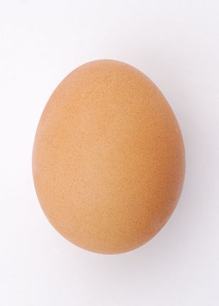
An ordinary chicken egg is the first think to come to mind for most people when they heard the word "egg". Photo by Sun Ladder, extracted from commons.wikimedia.org

An ordinary chicken egg is the first think to come to mind for most people when they heard the word “egg”. Photo by Sun Ladder*, extracted from commons.wikimedia.org

Eggs without a calcified shell rarely fossilize, and in fact there are no fossil eggs from Carboniferous, Permian and early Triassic, i.e., during all the history of non-mammalian synapsids.

But what’s so good in calcified shells? Well, a calcareous layer helps to prevent water loss from the embryo to the environment, whether to a reduction of relative humidity in the environment or a difference in temperature. Reptiles with non-calcified egg shells use to bury their eggs in damp soil where relative humidity approaches 100%, so dealing with the water loss problem. The increase of aridity from the end of the Permian to the Triassic may have favored the evolution of calcified eggs, but still there is no evidence of synapsid eggs. Could they have buried them in damp soil just as some reptiles do today? It’s possible, but…

Birds and mammals have developed a common feature: endothermy, i.e., “warm blood”. As we all know, birds use that to incubate eggs at elevated temperature, so speeding up the embryo development. It’s quite possible, and likely, that synapsids did the same. But as it was said, the difference in temperature between the egg and the environment also leads to water loss. In birds, the calcified shell solves this problem, but synapsids didn’t seem to have had this advantage.

Observing non-calcified eggs in snakes and lizards, it is possible to notice that they increase in mass by 10 to 100% after being laid. This happens due to water uptake from the environment and, as the shells is not rigid, the egg can also increase in volume, something that cannot happen in rigid-shelled eggs.

According to water uptake, eggs can be classified in endohydric and ectohydric. Endohydric eggs contain all water the embryo needs for the development when it is laid, while ectohydric eggs must take up water from the environment. In non-calcified eggs, it depends mainly of the eggs size. Large eggs may have enough water to sustain the embryo even with some water loss, since their surface:volume ratio is low, but the same cannot be applied to small eggs. And as it is known, the first mammals, and so their direct ancestors, were quite small creatures.

So what we have is an animal with non-calcified egg prone to dehydration and a high body temperature that can help to speed up the embryo development. How to join these two things?

The egg-laying mammal echidna and some marsupials have an abdominal pouch to retain eggs or newborns. It is known that the short-beaked echidnas incubate their eggs in the pouch. Female platypuses don’t have a pouch, but they incubate their eggs by curving their flat tail so that it touches the abdomen, forming a pouch-like enclosure. Inside this pouch, cutaneous secretions from the female’s abdomen would maintain the eggs moist, so preventing dehydration.

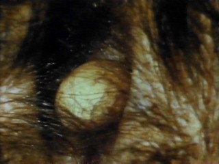
An egg of an echidna inside her pouch. (I found this picture on the internet without information about the author and stuff).

An egg of an echidna inside her pouch. (I found this picture on the internet without information about the author and stuff).

A problem in retaining eggs in a pouch is that it constrains the mobility of the parent. To solve this problem, eggs could stay some time inside the pouch to get hydrated and then be removed until it were necessary to rehydrate. This could let the female free from carrying the eggs along all the time and also increase the clutch size beyond the number of eggs that can fit in the pouch. This situation would have favored those females with more cutaneous secretions.

Once the young hatch from the eggs, they could also use the secretion as a source of food, which would lead to a progressive increase in the secretion’s nutritious value until it reached the status of milk.

It makes sense, doesn’t it? It does not contradict any kind of evidence about mammalian evolution and is based on true evidence through fossils and comparison with many kinds of extant groups.

– – –

References:

Oftedal, O. T. 2002. The origin of lactation as a water source for parchment-shelled eggs. Journal of mammary gland biology and neoplasia, 7 (3), 253-66 PMID: 12751890

Oftedal, O. T. 2002. The mammary gland and its origin during synapsid evolution. Journal of mammary gland biology and neoplasia, 7 (3), 225-52 PMID: 12751889

Creative Commons License
These works are licensed under a Creative Commons Attribution-ShareAlike 3.0 Unported License.

1 Comment

Filed under Evolution

Friday Fellow: Giant Tube Worm

by Piter Kehoma Boll

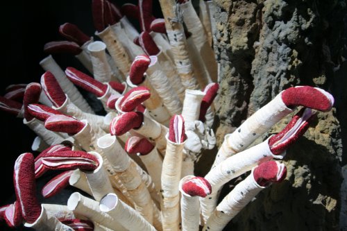
Giant tube worms Riftia pachyptila. Photo extrected from planeterde.de

Giant tube worms Riftia pachyptila. Photo extracted from planeterde.de

ResearchBlogging.org Let’s dive deep into the ocean and talk about this awesome animal, the giant tube worm Riftia pachyptila. Initially classified in a separate phylum, Vestimentifera, today it is included in a family of Annelids called Sibloginidae. Its common name comes from the fact that it can reach a length of 2.4 meters, quite big for a worm.

Endemic to deep-sea hydrothermal areas in the Pacific ocean, these worms are adapted to tolerate the high temperatures, pressure and levels of hydrogen sulfide in their environments. With their body protected by a chitin tube which can reach 3 meters in length, the only part exposed is a red structure, the branchial plume, highly vascularized ad rich in a hemoglobin complex of high molecular mass.

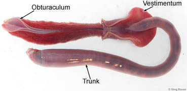
Below the plume lies the vestimentum, a muscular region which hosts the brain and the heart and is responsible for the extension and withdrawal of the plume. The name of the old phylum comprising this species, Vestimentifera, refers to this structure.

Follwing the vestimentum is the trunk and after it the opisthosome, which anchors the animal to the tube.

The plume is used to carry oxygen, carbon dioxide and sulfides into the animal’s body, which, however, lacks a mouth and gut.

A worm out of its tube. Photo extracted from spineless.ucsd.edu

A worm out of its tube. Photo extracted from spineless.ucsd.edu

To achieve nutrients, the giant tube worms host an endosymbiotic chemolithoautotrophic γ-Proteobacterium inside the trophosome, a richly vascularized organ in the trunk that constitutes a specific morphological adaptation to house the symbiotic bacteria. The sulfides are transported by the worm from the environment to the symbionts, which possess a sulfur oxidizing respiratory system and so can produce metabolic energy for themselves and for the worm.

The association between the giant tube worm and its chemoautrophic bacteria was the first of this kind to be described more than 30 years ago by Cavanaugh et al. and is currently the best studied one, but many questions about the details of this relationship, including the achievement of the bacteria by young worms, are yet to be fully answered.

Since the worm lacks a digestive system, its nutrition is entirely dependent on its symbiotic bacteria and all the anatomic adaptations designed to allow this association makes this a very good example of coevolution and make us think that there are no limits for life to adapt itself.

– – –

References:

Lopez-Garcia, P., Gaill, F., & Moreira, D. (2002). Wide bacterial diversity associated with tubes of the vent worm Riftia pachyptila. Environmental Microbiology, 4 (4), 204-215 DOI: 10.1046/j.1462-2920.2002.00286.x

Minic, Z., & Hervé, G. 2004. Biochemical and enzymological aspects of the symbiosis between the deep-sea tubeworm Riftia pachyptila and its bacterial endosymbiont. European Journal of Biochemistry, 271 (15), 3093-3102 DOI: 10.1111/j.1432-1033.2004.04248.x

Stewart FJ, & Cavanaugh CM 2006. Symbiosis of thioautotrophic bacteria with Riftia pachyptila. Progress in molecular and subcellular biology, 41, 197-225 PMID: 16623395

Leave a comment

Filed under Friday Fellow