Category Archives: cnidarians

Friday Fellow: Indo-Pacific Upsidedown Jellyfish

by Piter Kehoma Boll

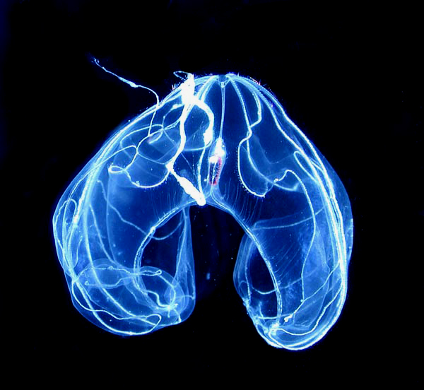
When we think of a jellyfish, we imagine it pulsating in the water column with its tentacles hanging from its underside. However some jellyfish do not behave exactly like that. This is the case, for example, with those of the genus Cassiopea, which includes the so-called upsidedown jellyfishes. The most studied species of Cassiopea is Cassiopea andromeda, which I decided to call the Indo-Pacific Upsidedown Jellyfish.

As you can imagine from the name I chose to it, the Indo-Pacific upsidedown jellyfish is native from the Indo-Pacific area. The reason why this and other species of this genus are called upsidedown jellyfish is because they are exactly that. They prefer not to swim around like regular a jellyfish, but rather stay near the bottom with their tentacles and mouth facing upward. As a result, they may end up being mistaken for sea anemones.

An adult Indo-Pacific upsidedown jellyfish measures up to 30 cm in diameter and has a brownish bell and arms with tentacles whose shape varies from pointy to flat, round or slender and the color varies from white to brown, red, pink, yellow, green or blue. This species is carnivorous, of course, like most cnidarians, and feeds on small animals that it captures and paralyzes.

The astonishing variety of colors and tentacle shapes in the Indo-Pacific upsidedown Jellyfish. Extracted from Lampert (2016).

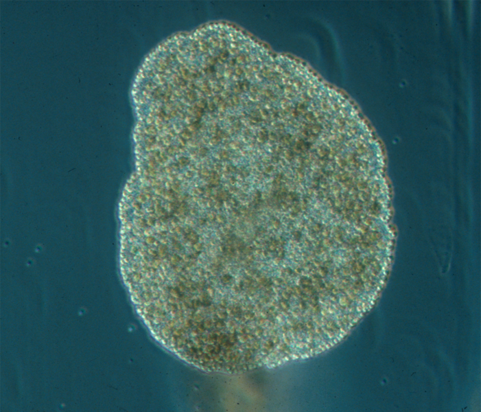
Adult Indo-Pacific upsidedown jellyfish are gonochoric, i.e., there are male and female specimens. Males release sperm in the water, which enters the body of the females and fertilizes the eggs. The eggs are kept inside the oral disc (the “mouth”) of the female until they develop into a free-living planula larva. The planula eventually settles when it founds a suitable substrate and develops into a polyp. The polyps can produce buds from their lower portion which detach and end up settling somewhere else to develop into new polyps. As the polyp grows, it turns into a young free-living medusa (ephyra), which grows to become and adult, restarting the cycle. An ideal place for the polyps to settle and develop are the roots of magroves, so this is one of the most common environments to find these jellyfish.

The Indo-Pacific upsidedown jellyfish is also known for its symbiotic relationship with zooxanthellae, unicellular algae (dinoflagellates) of the genus Symbiodinium. It is their presence that gives the jellyfish its brownish color. Both medusae and polyps have the algae inside them, but they are not passed vertically from the mother to the planulae. The polyps, instead, capture them freshly from the environment.

Specimens in an aquarium. Photo by Raimond Spekking.*

The algae provide nutrients for the jellyfish and the jellyfish, in turn, provides protection for the algae and ensures they will receive sunlight for photosynthesis. Several different Symbiodinium species are associated with the polyps, but this number often decreases to a single species in the adult medusae. As the shallow waters in which these jellyfish often live usually are prone to reach considerably high temperatures, not all zooxanthellae can survive.

Although the Indo-Pacific upsidedown jellyfish is native from the Indo-Pacific, it has been introduced in other parts of the world as well. It reached the Mediterranean probably through the Suez canal many decades ago and it has been recently found at the Atlantic coast of the Americas too. Another upsidedown jellyfish is native from this region, the Atlantic upsidedown jellyfish Cassiopea xamachana, and the consequences of both species meeting is still unknown. Let’s hope that the Indo-Pacific invader does not lead the Atlantic species to extinction.

– – –

Like us on Facebook!


Follow us on Twitter!

– – –

References:

Çevik, C., Erkol, I. L., & Toklu, B. (2006). A new record of an alien jellyfish from the Levantine coast of Turkey-Cassiopea andromeda (Forsskål, 1775)[Cnidaria: Scyphozoa: Rhizostomea]. Aquatic Invasions1(3), 196-197.

Hofmann, D. K., Neumann, R., & Henne, K. (1978). Strobilation, budding and initiation of scyphistoma morphogenesis in the rhizostome Cassiopea andromeda (Cnidaria: Scyphozoa). Marine Biology47(2), 161-176. https://doi.org/10.1007/BF00395637

Lampert, K. P. (2016). Cassiopea and its zooxanthellae. In The Cnidaria, past, present and future (pp. 415-423). Springer, Cham. https://doi.org/10.1007/978-3-319-31305-4_26

Morandini, A. C., Stampar, S. N., Maronna, M. M., & Da Silveira, F. L. (2017). All non-indigenous species were introduced recently? The case study of Cassiopea (Cnidaria: Scyphozoa) in Brazilian waters. Journal of the Marine Biological Association of the United Kingdom97(2), 321-328. https://repositorio.unesp.br/bitstream/handle/11449/162535/WOS000395463500012.pdf?sequence=1

Stampar, S. N., Gamero-Mora, E., Maronna, M. M., Fritscher, J. M., Oliveira, B. S., Sampaio, C. L., & Morandini, A. C. (2021). The puzzling occurrence of the upside-down jellyfish Cassiopea (Cnidaria: Scyphozoa) along the Brazilian coast: a result of several invasion events?. Zoologia (Curitiba)37. https://doi.org/10.3897/zoologia.37.e50834

– – –

*Creative Commons License This work is licensed under a Creative Commons Attribution-ShareAlike 4.0 International License.

Leave a comment

Filed under cnidarians, Conservation, Friday Fellow

Friday Fellow: Lion’s Mane Jellyfish

by Piter Kehoma Boll

Today is time to talk again about a celebrity.

Although three cnidarians have been featured as Friday Fellows already, none of them was a jellyfish until now. So let’s introduce the first one and let it be a considerably famous species, the lion’s mane jellyfish Cyanea capillata.

Lion’s mane jellyfish near Newfoundland, Canada. Credits to Derek Keats.**

The lion’s mane jellyfish is a very large jellyfish, among the largest species known to date. Its bell can reach up to 2 m in diameter and its tentacles can grow up to 30 m in length, thus becoming longer than a blue whale! It is an inhabitant of the very cold waters of the Arctic and neighboring areas of the Atlantic and Pacific oceans. It cannot survive in warm waters and specimens living in the southernmost areas of its range cannot even grow to the full size.

The color of an adult lion’s mane jellyfish is pale red or pale yellow. Its genus name, Cyanea, which refers to a blue color, is due to another species of the genus, the blue jellyfish, Cyanea lamarckii. Its specific epithet, capillata as well as its common name are references to its dense mass of tentacles that resemble a lion’s mane. The jellyfish’s bell is divided into eight lobes, each lobe having from 70 to 150 tentacles. The indentations between the lobes contain a special organ, the rhopalium, that helps jellyfishes orient themselves in water.

A specimen in Norway. Credits to Arstein Rønning.*

Like all jellyfishes, the lion mane jellyfish has a complex life cycle. Adult specimens reproduce sexually in summer, with males releasing sperm into the water. The sperm swims into the body of the female, where the eggs are fertilized. The first life stage, the larva, grows inside the body of the female and is then released into the water where it attaches to a surface to become a polyp. The larvae seem to prefer rougher surfaces to attach and especially in darker places. The polyp grows during winter and reproduces asexually during spring. The asexual reproduction, called strobilation, occurs by the polyp releasing segments that become ephyrae, which are like very young jellyfish. The ephyrae grow to become adult jellyfish and restart the cycle.

The lion’s mane jellyfish feeds on a great variety of species during its life cycle, including plankton, invertebrates and even small vertebrates. Nevertheless, its huge body is also used as the habitat of several other animals that live in the cold northern waters. It also has a complicated relationship with the moon jellyfish, Aurelia aurita, with which it shares its habitat. While adult lion’s mane jellyfish prey on adult moon jellyfish, adult moon jellyfish prey on larvae and ephyrae of the lion’s mane jellyfish, and both species also compete for the same prey.

A lion’s mane jellyfish (top-right) capturing a moon jellyfish (bottom-left). Photo by W. Carter.

Like all cnidarians, the lion’s mane jellyfish stings. The contat with a single tentacle in humans usually does not cause much complication except for those with some sort of allergy or sensitivity. However, if you are unfortunate enough to end up swimming directy into the tentacle mass of a specimen, becoming covered by that stinging nightmare, you may end up having to be taken to a hospital quickly. Despite the low risk of killing a human, one lion’s mane jellyfish became famous as the assassin in one of Sherlock Holmes’ cases. The victim was an unfortunate guy with a heart condition, though.

– – –

More cnidarians:

Friday Fellow: Deep-Sea Marr (on 22 April 2016)

Friday Fellow: Portuguese Man o’ War (on 7 July 2017)

Friday Fellow: Blue Coral (on 18 May 2018)

– – –

Like us on Facebook!

Follow us on Twitter!

– – –

References:

Brewer RH (1976) Larval settling behavior of Cyanea capillata (Cnidaria: Scyphozoa). The Biological Bulletin 150(2). https://doi.org/10.2307/1540467

Gröndahl F, Hernroth L (1987) Release and growth of Cyanea capillata (L.) ephyrae in the Gullmar Fjord, western Sweden. Journal of Experimental Marine Biology and Ecology 106(1):91–101. https://doi.org/10.1016/0022-0981(87)90149-3

Gröndahl F (1988) A comparative ecological study on the scyphozoans Aurelia aurita, Cyanea capillata and C. lamarckii in the Gullmar Fjord, western Sweden, 1982 to 1986. Marine Biology 97: 541–550. https://doi.org/10.1007/BF00391050

Wikipedia. Lion’s mane jellyfish. Available at < https://en.wikipedia.org/wiki/Lion%27s_mane_jellyfish >. Access on 3 September, 2020.

– – –

*Creative Commons License This work is licensed under a Creative Commons Attribution-ShareAlike 3.0 Unported License.

**Creative Commons License This work is licensed under a Creative Commons Attribution-Share Alike 2.0 Generic License.

Leave a comment

Filed under cnidarians, Friday Fellow

Friday Fellow: Blue Coral

by Piter Kehoma Boll

Sorry, guys! It has been about three weeks since my last post, but I was too busy with a lot of personal and academic stuff and wasn’t able to dedicate any time to the blog, but I’m back!

Let’s return with a marine animal as today’s Friday Fellow, the called blue coral, Heliopora coerulea.

heliopora_coerulea2c_koh_phangan

A colony of the blue coral in Thailand. Credits to Chaloklum Diving.*

Found in tropical waters of the Pacific and Indian oceans, the blue coral is a peculiar species, being the only one in the genus Heliopora and in the family Helioporidae. It is the only species in the subclass Octocorallia that has a massive skeleton, a feature more common in the stony corals of the subclass Hexacorallia. As a result, the ecological role of the blue coral is usually closer to that of stony corals that to that of its closer relatives.

The skeleton of the blue coral is composed of aragonite and has a distinctive bluish-gray color caused by the presence of iron salts. There are fossils of bluish corals with the same morphology that date back to the Cretaceous, indicating that this is a very old species.

450px-blue_coral_-_heliopora_coerulea

A skeleton of the blue coral in the Natural History Museum, London. Photo by Wikimedia user Kinkreet.**

Although widespread, the blue coral is currently considered a vulnerable species, with some population showing very low genetic diversity. This species is threatened mainly by the jewelry and aquarium trades and by the acidification of the oceans.

– – –

Like us on Facebook!

Follow us on Twitter!

– – –

References:

Babcock, R. (1990) Reproduction and development of the blue coral Heliopora coerulea (Alcyonaria: Coenothecalia)Marine Biology 104: 475–481.

EOL: Encyclopedia of Life. Heliopora coerulea. Available at < http://eol.org/pages/1006937/overview >. Access on May 14, 2018.

Wikipedia. Heliopora coerulea. Available at < https://en.wikipedia.org/wiki/Blue_coral >. Access on May 14, 2018.

Yasuda, N.; Taquet, C.; Nagai, S.; Fortes, M.; Fan, T.-Y.; Phongsuwan, N.; Nadaoka, K. (2014) Genetic structure and cryptic speciation in the threatened reef-building coral Heliopora coerulea along Kuroshio Current. Bulletin of Marine Science 90(1): 233–255. https://doi.org/10.5343/bms.2012.1105

– – –

*Creative Commons License
This work is licensed under a Creative Commons Attribution 3.0 Unported License.

**Creative Commons License
This work is licensed under a Creative Commons Attribution-ShareAlike 3.0 Unported License.

1 Comment

Filed under cnidarians, Friday Fellow, Zoology

Friday Fellow: Portuguese Man o’ War

by Piter Kehoma Boll

And so we finally reached the 100th Friday Fellow! In order to commemorate, we will have two Friday Fellows today, just as we had during the 50th one. And to start I chose a cnidarian that always caught me attention.

Living in the Atlantic Ocean and known popularly as Portuguese man o’ war, its binomial name is Physalia physalis, both words derived from the Greek word for bubble, physalis. And the Portuguese man o’ war is, in fact, like a floating bubble with some stuff attached, or at least it looks like that.

Physalia_physalis2

A Portuguese man o’ war lying on the beach. Photo by Anna Hesser.*

Most people may think that the Portuguese man o’ war is a jellyfish due to its looks, but it is actually part of another group of cnidarians, the siphonophores. Their body is not a single individual, but rather a colony of several smaller animals, called zooids, which are speciallized to have different functions within the colony and cannot live separately. They are all derived from the same embryo, thus being clones from each other.

The upper portion of the Portuguese man o’ war has a gas-filled sack, which is called the pneumatophore and is the original organism derived directly from the embryo. Below the pneumatophore there are several different kinds of organisms, such as nectophores for swimming, dactylozooids for defense and capture of prey, gonozooid for reproduction and gastrozooids for feeding. The long tentacles, which reach more than 10 m in length, are composed by dactylozooids and fish for prey throughout the water.

Physalia_physalis1

Floating on the sea. Photo by Regine Stiller.*

As other cnidarians, the Portuguese man o’ war has nettle-like cells which sting and inject venom. In humans, the venom usually cause pain and let whip-like marks on the skin where the tentacles touched. Sometimes more severe complications will results and in rare cases it may result in death.

– – –

References:

Stein, M. R.; Marraccini, J. V.; Rothschild, N. E.; Burnett, J. W. (1989) Fatal portuguese man-o’-war (Physalia physalis) envenomation. Annals of Emergency Medicine 18(3): 312–315.

Wikipedia. Portuguese man o’ war. Available at <https://en.wikipedia.org/wiki/Portuguese_man_o%27_war&gt;. Access on June 16, 2017.

– – –

*Creative Commons License
This work is licensed under a Creative Commons Attribution-NonCommerical-NoDerivs 2.0 Generic License.

1 Comment

Filed under cnidarians, Friday Fellow

Who came first? The comb or the sponge?

by Piter Kehoma Boll

The endless question is here again, but this time it appears to be settled. What animal group is the earliest of all? Who came first?

It is clear that there are five animal lineages that are usually regarded as monophyletic: sponges, placozoans, comb jellies, cnidarians and bilaterians. Let’s take a brief look at each of them:

Sponges (phylum Porifera) are always sessile, i.e., they do not move and are fixed to the substrate. They have a very simple anatomical structure. Their body is consisted of a kind of tube, having a large internal cavity and two layers of cells, an outer one and an inner one around the cavity. There are several small openings connecting the cavity to the outside, called pores, and one or more large cavities, called oscula (singular: osculum). Between the two cell layers there is a jelly-like mesohyl containing unspecialized cells, as well as the skeleton structures, including fibers of spongine and spicules of calcium carbonate or silica. Some species also secrete an outer calcium carbonate skeleton over which the organic part grows. Sponges lack muscles, nervous system, excretory system or any other kind of system. They simply live by beating the flagella of the choanocytes (the cells of the inner layer), creating a water flow entering through the pores and exiting through the osculum. The choanocytes capture organic particles in the water and ingest them by phagocytosis. All sponge cells can change from one type to another and migrate from one layer to another, so there are no true tissues.

porifera_body_structures_01

Body structures found in sponges. Picture by Philip Chalmers.*

Placozoans (phylum Placozoa) are even simpler than sponges, but they actually have true tissues. They are flat amoeboid organisms with two layers of epithelium, one dorsal and one ventral, and a thin layer of stellate cells. The ventral cell layer is slightly concave and appears to be homologous to the endoderm (the “gut” layer) of other animals, while the upper layer is homologous to the ectoderm (the “skin” layer).

701px-trichoplax_adhaerens_photograph

Trichoplax adhaerens, the only species currently in the phylum Placozoa. Photo by Bernd Schierwater.**

Comb jellies (phylum Ctenophora) resemble jellyfishes, but a closer look reveals many differences. Externally they have an epidermis composed by two layers, an outer one that contains sensory cells, mucus-secreting cells and some specialized cells, like colloblasts that help capturing prey and cells containing multiple cilia used in locomotion, and an inner layer with a nerve net and muscle-like cells. They have a true mouth that leads to a pharynx and a stomach. From the stomach, a system os channels distribute the nutrients along the body. Opposite to the mouth there is a small anal pore that may excrete small unwanted particles, although most of the rejected material is expelled through the mouth. There is a layer of jelly-like material (mesoglea) between the gut and the epidermis.

bathocyroe_fosteri

The comb jelly Bathocyroe fosteri.

Cnidarians (phylum Cnidaria) have a structure similar to comb jellies, but not as complex. They also have an outer epidermis, but this is composed by a single layer of cells, and a sac-like gut surrounded by epthelial cells (gastrodermis), as well as a mesoglea between the two. Around the mouth there is one or two sets of tentacles. The most distinguishing feature of cnidarians is the presence of harpoon-like nettle cells, the cnidocytes, which are used as a defense mechanism and to help subdue prey.

800px-cross_section_jellyfish_en-svg

Body structure of a cnidarian (jellyfish). Picture by Mariana Ruiz Villarreal.

Bilaterians (clade Bilateria) includes all other animals. They are far more complex and are characterized by a bilateral body, cephalization (they have heads) and three main cell layers, the ectoderm, which originates the epidermis and the nervous system, the mesoderm, which give rise to muscles and blood cells, and the endoderm, which develops into the digestive and endocrine systems.

500px-bilaterian-plan-svg

Basic bilaterian structure.

Traditionally, sponges were always seen as the most primitive animals due to their lack of true tissues, muscular cells, nervous cells and all that stuff. However, some recent molecular studies have put the comb jellies as the most primitive animals. This was highly unexpected, as comb jellies are far more complex than sponges and placozoans, which would suggest that muscles and a nervous system evolved twice in the animal kingdom, or that sponges are some weird simplification of a more complex ancestor, which would be very hard to explain. The nervous system of comb jellies is indeed quite unusual, but not so much that it needs an independent origin.

However, now things appear to be settled. A study published this month on Current Biology by Simion et al. reconstructed a phylogenetic tree using 1719 genes of 97 animal species, and applying new and more congruent methods. With this more refined dataset, they recovered the classical reconstruction that puts sponges at the base of the animal tree, a more plausible scenario after all.

But why other studies have found comb jellies as the most basal group? Well, it seeems that comb jellies have unusually high substitution rates, meaning that their genes evolve faster. This leads to a problem called “long branch attraction” in phylogenetic reconstructions. As DNA has only four different nucleobases, namely adenine, guanine, cytosine and thymine, each one can only mutate into one of the other three. When mutations occur very often, they may go back to what they were in long lost ancestor, leading to misinterpretations in the evolutionary relationships. That seems to be what happens with comb jellies.

So, it seems that after all the sponge indeed came first.

– – –

References and further reading:

Borowiec ML, Lee EK, Chiu JC, & Plachetzki DC 2015. Extracting phylogenetic signal and accounting for bias in whole-genome data sets supports the Ctenophora as sister to remaining Metazoa. BMC Genomics 16: 987. DOI: 10.1186/s12864-015-2146-4

Littlewood DTJ 2017. Animal Evolution: Last Word on Sponges-First? Current Biology 27: R259–R261. DOI: 10.1016/j.cub.2017.02.042

Simion P, Philippe H, Baurain D, Jager M, Richter DJ, Di Franco A, Roure B, Satoh N, Quéinnec É, Ereskovsky A, Lapébie P, Corre E, Delsuc F, King N, Wörheide G, & Manuel M 2017. A Large and Consistent Phylogenomic Dataset Supports Sponges as the Sister Group to All Other Animals. Current Biology 27: 958–967. DOI: 10.1016/j.cub.2017.02.031

Wallberg A, Thollesson M, Farris JS, & Jondelius U 2004. The phylogenetic position of the comb jellies (Ctenophora) and the importance of taxonomic sampling. Cladistics 20: 558–578. DOI: 10.1111/j.1096-0031.2004.00041.x
– – –
Creative Commons License
This work is licensed under a Creative Commons Attribution-ShareAlike 3.0 Unported License.
** Creative Commons License
This work is licensed under a Creative Commons Attribution 4.0 International License.

Leave a comment

Filed under cnidarians, Evolution, sponges, Zoology

Friday Fellow: Deep-sea marr

by Piter Kehoma Boll

Looking like some sort of tribal mystic rattle, our newest Friday Fellow comes from the deep waters in the northern hemisphere. Its scientific name is Marrus orthocanna, and I adapted a common name as “deep-sea marr”. I think it sounds cool.

A magic rattle form the deep sea. Photo by Kevin Raskoff

A magic rattle from the deep sea. Photo by Kevin Raskoff.

The deep-sea marr is a siphonophore cnidarian, and as all siphonophores, it is a colonial organism rather than a single individual. It is composed by several specialized organisms (zooids) linked together by a long “stem” and unable to live independently. It is a free-swimming organism, swiming in a pulsative way through the dark deep sea waters.

At the front or upper side of the colony is the pneumatophore, a gas-filled float that is the primordial organism in the colony, the one that originated directly from the embryo. After it, there are several bell-shaped translucent organisms, the nectophores, which are specialized in locomotion, making the colony move by contractions. The last part of the body, the siphosome, contains a series of different zooids, including individuals specialized to capture prey, to digest food and to reproduce.

The deep-sea marr is found mainly in Arctic waters, but sometimes occurs more southwards, down to the Mediterranean Sea. It may grow to several meters in length and its diet most likely includes small crustaceans. It is a weird, but certainly beautiful creature.

– – –

References:

EOL –  Encyclopedia of Life: Marrus orthocanna. Available at: < http://eol.org/pages/1005745 >. Access on April 21, 2016.

Wikipedia. Marrus orthocanna. Available at: <https://en.wikipedia.org/wiki/Marrus_orthocanna >. Access on April 21, 2016.

2 Comments

Filed under cnidarians, Friday Fellow, Zoology