Monthly Archives: November 2017

Friday Fellow: Chinese Magnolia Vine

by Piter Kehoma Boll

Coming from the forests of Northern China, Korea and Eastern Russia, our newest fellow is a woody vine called Schisandra chinensis and populary known as Chinese magnolia vine.

394px-schisandra_chinensis_2

The beautiful red fruits of the Chinese magnolia vine. Photo by Vladimir Kosolapov.*

Used in Chinese traditional medicine, the plant is considered one of the 50 fundamental herbs. The part of the plant most commonly used are the berries, which are known as magnolia berries or five-flavor-fruits. The second name is a translation of the Chinese name, 五味子 (wǔwèizi), because the berry is said to contain all five basic Chinese flavors: salty, sweet, sour, spicy and bitter. An infusion prepared with the dried fruits is called omija tea or omija-cha, from the Korean name of the fruits.

800px-omija-cha

A cup of omija tea. Photo by Raheel Shahid.**

The traditional uses of the Chinese magnolia vine included the treatment of disorders related mainly to the sexual organs. Several current studies by laboratory trials indicated that the plant has a large number of beneficial properties, including antioxidant properties and the ability to increase endurance, working ability, accuracy of movements and mental ability. It also seems to be useful in the treatment of several diseases and disorders, especially inflamatory ones, such as sinusitis, otitis, neuritis, dermatitis and gastritis, as well as on some infectious diseases such as influenza and pneumonia, among many other conditions.

I’m certainly interested in trying a cup of omija tea. What about you? Have you ever had the chance?

– – –

Likes us on Facebook!

– – –

References:

Panossian, A.; Wikman, G. (2008) Pharmacology of Schisandra chinensis Bail.: An overview of Russian research and uses in medicine. Journal of Ethnopharmacology 118(2): 183-212. https://doi.org/10.1016/j.jep.2008.04.020

Wikipedia. Schisandra chinensis. Available at < https://en.wikipedia.org/wiki/Schisandra_chinensis >. Access on October 31, 2017.

– – –

*Creative Commons License
This work is licensed under a Creative Commons Attribution 3.0 Unported License.

**Creative Commons License
This work is licensed under a Creative Commons Attribution 2.0 Generic License.

1 Comment

Filed under Botany, Friday Fellow

Friday Fellow: Brown Mussel

by Piter Kehoma Boll

Until now, the mollusks featured here included a chiton, a cephalopod and two gastropods. So it is time to bring a bivalve. And what would be better than showing you a common mollusk from the South Atlantic Ocean?

Living on rocky shores around South America and Africa, our fellow is called Perna perna, or populary brown mussel. In places where it lives, it can be found in great concentrations, sometimes covering large areas of rocks. It usually measures about 90 mm in length, but some larger specimens may reach up to 120 mm. The increased surface area on the rocks they occupy attract other rock-living marine species, such as barnacles, limpets, snails and algae.

Perna_perna

Some specimens of Perna perna growing on an oyster in South Africa. Photo by Bernadette Hubbart.*

The brown mussel is a filter feeder, as most bivalves, feeding on suspended organic matter, as well as on small microrganisms, such as phytoplankton and zooplankton. As a prey, it is eaten by a variety of animals, such as sea birds, crustaceans and mollusks. Humans also consume it in both South America and Africa. Its ingestion, however, must be cautious, as it may contain toxins from dinoflagellates that it ingested, as well as heavy metals from water pollutants.

Spread through the world by humans after attaching itself on ships, the brown mussel has become invasive in other parts, especially in the Gulf of Mexico, and it continues to increase its occupied area. This can have deleterious effects both ecologically and economically, as it may displace native species and also cause damage to human equipments. It is, therefore, one more species that became a problem due to us, humans. And the damage will not be easy to be repared.

– – –

Likes us on Facebook!

– – –

References:

Ferreira, A. G.; Machado, A. L. S.; Zalmon, I. R. (2004) Temporal and spatial variation on heavy metal concentrations in the bivalve Perna perna (LINNAEUS, 1758) on the northern coast of Rio de Janeiro State, Brazil. Brazilian Archives of Biology and Technology 47(2): 319–327. http://dx.doi.org/10.1590/S1516-89132004000200020

Holland, B. S. (2001) Invasion without a bottleneck: microsatellite variation in natural and invasive populations of the brown mussel Perna perna (L). Marine Biotechnology 3, 407–415. https://dx.doi.org/10.1007/s1012601-0060-Z

Wikipedia. Perna perna. Available at: < https://en.wikipedia.org/wiki/Perna_perna >. Access on October 21, 2017.

– – –

*Creative Commons License
This work is licensed under a Creative Commons Attribution-NoDerivs 2.0 Generic License.

1 Comment

Filed under Friday Fellow, mollusks, Zoology

Friday Fellow: Yellow Morel

by Piter Kehoma Boll

Time for our next fungus, and this time it is a delicious one, or at least I think so, as I have never eaten it. Scientifically known as Morchella esculenta, its common names include common morel, yellow morel, true morel or simply morel.

576px-old_holiday_shot_of_morchella_esculenta_28gb3d_morel_mushroom2c_d3d_speise-morchel2c_nl3d_gewone_morielje29_-_panoramio

A fruiting body of the yellow morel in France. Photo by Henk Monster.*

Common in North America and Europe, as well as in some parts of Asia, especially in wooden areas, the yellow morel is a popular edible fungus of the phylum Ascomycota, so it is not closely related to the more common mushrooms, but it is a relative of the truffles, for example.

Morels are usually easily recognizable due to their peculiar appearance. Appearing during spring, their fruiting body is more or less oval in shape, covered with irregular pits and ridges, and hollow.

450px-morchella-esculenta-001

An open morel showing its hollowness. Photo by Wikimedia user 00Amanita00.*

Although being one of the most highly prized mushrooms, morels can give you some undesirable effects, such as gastrointestinal problems, if eaten raw or if too old. So, it is advisable to eat young mushrooms and at least blanching them before consumption. As they are hollow, it is common to eat them stuffed with vegetables or meat.

Pharmacological and biochemical studies revealed that the yellow morel has many healthy properties, such as the presence of antioxidants and substances that stimulate the immune system, as well as anti-inflammatory and antitumour properties. It is certainly a food that is worth to include in our diet, too bad that is tends to be kind of expensive…

– – –

Like us on Facebook!

– – –

References:

Duncan, C. J. G.; Pugh, N.; Pasco, D. S.; Ross, S. A. (2002) Isolation of galactomannan that enhances macrophage activation from the edible fungs Morchella esculentaJournal of Agricultural and Food Chemistry, 50(20): 5683–5695. DOI: 10.1021/jf020267c

Mau, J.-L.; Chang, C.-N.; Huang, S.-J.; Chen, C.-C. (2004) Antioxidant properties of methanolic extracts from Grifola frondosa, Morchella esculenta and Termitomyces albuminosus mycelia. Food Chemistry, 87(1): 111-118.
https://doi.org/10.1016/j.foodchem.2003.10.026

Nitha, B.; Meera, C. R.; Janardhanan, K. K. (2007) Anti-inflammatory and antitumour activities of cultured mycelium of morel mushroom, Morchella esculentaCurrent Science, 92(2): 235–239.

Wikipedia. Morchella esculenta. Available at < https://en.wikipedia.org/wiki/Morchella_esculenta >. Access on October 31, 2017.

– – –

*Creative Commons License
This work is licensed under a Creative Commons Attribution 3.0 Unported License.

3 Comments

Filed under Friday Fellow, Fungi

The hammerhead flatworms: once a mess, now even messier

by Piter Kehoma Boll

Few people know that land planarians exist, but when they do, they most likely know the hammerhead flatworms, which comprise the subfamily Bipaliinae.

The hammerhead flatworms, or simply hammerhead worms, have this name because their head has lateral expansions that make them resemble a hammer, a shovel or a pickaxe. Take a look:

Bipalium_vagum

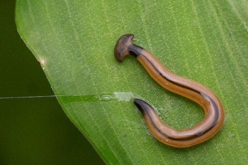
The “wandering hammerhead worm”, Bipalium vagum. Notice the peculiar head. Photo by flickr user budak.*

The Chinese knew the hammerhead worms at least since the 10th century, which is understandable, since they are distributed from Japan to Madagascar, including all southern and southeast Asia, as well as Indonesia, the Philippines and other archipelagos. The western world, however, first heard of them in 1857, when William Stimpson described the first species and put them in a genus called Bipalium, from Latin bi- (two) + pala (shovel), due to the head shape. One of them was the species Bipalium fuscatum, a Japanese species that is currently considered the type species of the genus.

800px-bipalium_fuscatum_by_head

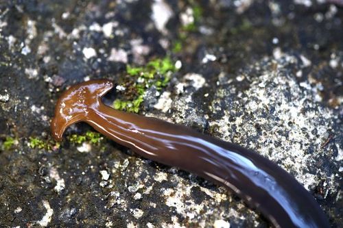
Anterior region of Bipalium fuscatum, the “brownish hammerhead worm”. Photo by Wikimedia user 根川大橋.**

Two years later, in 1859, Ludwig K. Schmarda described one more species, this one from Sri Lanka, and, unaware of Stimpson’s paper, called the species Sphyrocephalus dendrophilus, erecting the new genus for it from Greek sphȳra (hammer) + kephalē (head).

Sphyrocephalus_dendrophilus

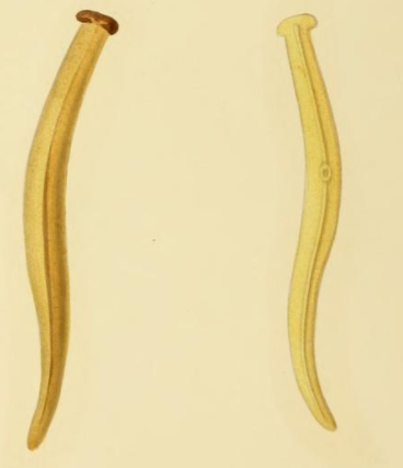
Drawings by Schmarda of Sphyrocephalus dendrophilus.

In the next year, 1860, Edward P. Wright did something similar and described some hammerhead worms from India and China, creating a new genus, Dunlopea, for them. The name was a homage to his friend A. Dunlop (whoever he was).

Dunlopea_grayia

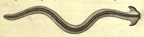
Wright’s Drawing of Dunlopea grayia (now Diversibipalium grayi) from China.

Eventually those errors were perceived and all species were put in the genus Bipalium, along with several others described in the following years. All hammerhead worms were part of the genus Bipalium until 1896, when Ludwig von Graff decided to improve the classification and divided them into three genera:

1. Bipalium: With a head having long “ears”, a well developed head.
2. Placocephalus (“plate head”): With a more semicircular head.
3. Perocephalus (“mutilated head”): With a shorter, rudimentary head, almost as if it had been cut off.

Bipaliids

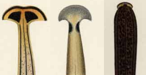
Compare the heads of typical species of Bipalium (left), Placocephalus (center) and Perocephalus (right), according to Graff.

This system, however, was soon abandoned and everything went back to be simply Bipalium and continued that way for almost a century, changing again only in 1998, when Kawakatsu and his friends started to mess with the penises of the hammerhead worms.

First, in 1998, they erected the genus Novibipalium (“new Bipalium“) for species with a reduced or absent penis papilla, and retained in Bipalium those with a “well”-developed penis papilla. It is worth noticing though that this well-developed papilla is not much bigger than a reduced papilla in Novibipalium. In both genera the actual, functional penis is formed by a set of folds in the male atrium and not by the penis papilla itself as in other land planarians that have a penis papilla.

Later, in 2001, Ogren & Sluys separated some more species of Bipalium in a new genus called Humbertium (after Aloïs Humbert, who described most species of this new genus). They were separated from Bipalium because the ovovitelloducts (the ducts that conduct the eggs and vitellocites) enter the female atrium from ahead, and not from behind as in the typical Bipalium. This separation is, in my opinion, more reasonable than the previous one.

Now we had three genera of hammerhead worms based on their internal anatomy, but several species were described without any knowledge of their sexual organs. Thus, in 2002, Kawakatsu and his friends created one more genus, Diversibipalium (the “diverse Bipalium“) to include all species whose anatomy of the sexual organs was unknown. In other words, it is a “wastebasket” genus to place them until they are better studied.

Are these three genera, Bipalium, Novibipalium and Humbertium, as now defined, natural? We still don’t know, but I bet they are not. A good way to check it would be by using molecular phylogeny, but we don’t have people working with these animals in their natural habitats, so we do not have available material for that. Another thing that can give us a hint is to look at their geographical distribution. We can assume that genetically similar species, especially of organisms with such a low dispersal ability as land planarians, all occur in the same geographical region, right? So where do we find species of each genus? Let’s see:

Bipalium: Indonesia, Japan, China, Korea, India.

Novibipalium: Japan.

Humbertium: Madagascar, Sri Lanka, Southern India, Indonesia.

Weird, right? They are completely mixed and covering a huge area of the planet, especially when we consider Humbertium. We can see a tendency, but nothing very clear.

Fortunately, some molecular analyses were published (see Mazza et al. (2016) in the references). One, which included the species Bipalium kewense, B. nobile, B. adventitium, Novibipalium venosum and Diversibipalium multilineatum placed Diversibipalium multilineatum very close to Bipalium nobile, and they are in fact very similar, so I guess that we can transfer it from Diversibipalium to Bipalium, right? Similary, Novibipalium venosum appears mixed with the species of Bipalium. I guess this is kind of messing things up one more time.

681px-bipalia_invasive

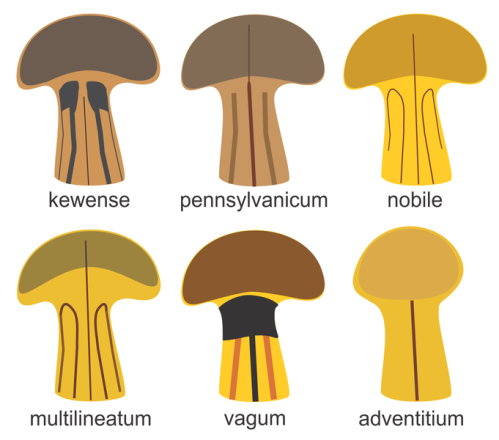
Head of some species of Bipalium, including the ones used in the study cited above. Unfortunately, I couldn’t find a photo or drawing of Novibipalium venosum. Image by myself, Piter Kehoma Boll.**

Interestingly, among the analyzed species, the most divergent was Bipalium adventitium, whose head is “blunter” than that of the other ones. Could the head be the answer, afterall? Let’s hope that someone with the necessary resources is willing to solve this mess soon.

– – –

Like us on Facebook!

– – –

See also:

Once found and then forgotten: the not-so-bright side of taxonomy.

The lack of taxonomists and its consequences on ecology.

They only care if you are cute. How charisma harms biodiversity.

The faboulous taxonomic adventure of the genus Geoplana.

Darwin’s Planaria elegans: hidden, extinct or misidentified?

– – –

References:

Graff, L. v. (1896) Über das System und die geographische Verbreitung der Landplanarien. Verhandlungen der Deutschen Zoologischen Gesellschaft6: 61–75.

Graff, L. v. (1899) Monographie der Turbellarien. II. Tricladida Terricola (Landplanarien). Engelmann, Leipzig.

Kawakatsu, M.; Ogren, R. E.; Froehlich, E. M. (1998) The taxonomic revision of several homonyms in the genus Bipalium, family Bipaliidae (Turbellaria, Seriata, Tricladida, Terricola). The Bulletin of Fuji Women’s College Series 236: 83–93.

Kawakatsu, M.; Ogren, R. E.; Froehlich, E. M., Sasaki, G.-Y. (2002) Additions and corrections of the previous land planarians indices of the world (Turbellaria, Seriata, Tricladida, Terricola). The bulletin of Fuji Women’s University. Ser. II40: 162–177.

Mazza, G.; Menchetti, M.; Sluys, R.; Solà, E.; Riutort, M.; Tricarico, E.; Justine, J.-L.; Cavigioli, L.; Mori, E. (2016) First report of the land planarian Diversibipalium multilineatum (Makino & Shirasawa, 1983) (Platyhelminthes, Tricladida, Continenticola) in Europe. Zootaxa4067(5): 577–580.

Ogren, R. E.; Sluys, R. (2001) The genus Humbertium gen. nov., a new taxon of the land planarian family Bipaliidae (Tricladida, Terricola). Belgian Journal of Zoology131: 201–204.

Schmarda, L. K. (1859) Neue Wirbellose Thiere beobachtet und gesammelt auf einer Reise um die Erde 1853 bis 1857 1. Turbellarien, Rotatorien und Anneliden. Erste Hälfte. Wilhelm Engelmann, Leipzig.

Stimpson, W. (1857) Prodromus descriptionis animalium evertebratorum quæ in Expeditione ad Oceanum, Pacificum Septentrionalem a Republica Federata missa, Johanne Rodgers Duce, observavit er descripsit. Pars I. Turbellaria Dendrocœla. Proceedings of the Academy of Natural Sciences of Philadelphia9: 19–31.

Wright, E. P. (1860) Notes on Dunlopea. Annals and Magazine of Natural History, 3rd ser.6: 54–56.

– – –

*Creative Commons License
This work is licensed under a Creative Commons Attribution-NonCommerical-NoDerivs 2.0 Generic License.

**Creative Commons License
This work is licensed under a Creative Commons Attribution-ShareAlike 4.0 International License.

2 Comments

Filed under flatworms, Systematics, taxonomy, worms, Zoology

Friday Fellow: Common Latticed Sponge

by Piter Kehoma Boll

Let’s go back to the sea and to our distant animal relatives, the sponges. Today I’m bringing a calcareous sponge with a nice appearance, Clathrina clathrus, who I decided to call “the common latticed sponge”.

Found in the Mediterranean Sea and the European coast of the Atlantic Ocean, the common latticed sponge has a yellow color and about 10 cm in diameter. It is formed by a tangle of tubes that somewhat resemble a twisted lattice or something like that.

450px-clathrina_clathrus_scarpone_055

A specimen of Clathrina clathrus with its latticed appearance. Photo by Wikimedia user Esculapio.*

The shape and size of the specimens is quite variable, changing in a matter of hours by expansion, contraction and folding of structures and cells. In the same way, specimens often fragment into smaller ones or merge into larger ones, so that individuality is a dynamic process.

Recently, the common latticed sponge has revealed to contain some compounds, known as clathridimines, that show antimicrobial activities against Gram-positive and Gram-negative bacteria, as well as against the yeast Candida albicans. These compounds may be produced by the diverse community of bacteria that live in close association with this sponge, a community that is yet very little known.

– – –

Like us on Facebook!

– – –

References:

Gaino, E.; Pansini, M.; Pronzato, R.; Cicogna, F. (1991) Morphological and structural variations in Clathrina clathrus (Porifera, Calcispongiae). In.: Reitner, J.; Keupp, H. (Eds.) Fossil and Recent Sponges. Springer-Verlag, Berlin. pp. 360-371.

Quévrain, E.; Roué, M.; Domart-Coulon, I.; Bourguet-Kondracki, M.-L. (2014) Assessing the potential bacterial origin of the chemical diversity in calcareous sponges. Journal of Marine Science and Technology 22(1): 36-49.

Roué, M.; Domart-Coulon, I.; Ereskovsky, A.; Djediat, C.; Perez, T.; Bourguet-Kondracki, M.-L. (2010) Cellular localization of clathridimine, an antimicrobial 2-aminoimidazole alkaloid produced by the Mediterranean calcerous sponge Clathrina clathrusThe Journal of Natural Products 73(7): 1277–1282.

– – –

*Creative Commons License
This work is licensed under a Creative Commons Attribution 3.0 Unported License.

Leave a comment

Filed under Friday Fellow, sponges