Category Archives: Parasites

Friday Fellow: Mediterranean Plumefoot Mite

by Piter Kehoma Boll

There are endless forms of beauty among the small creatures that we often do not see around us. Mites, which are so ubiquitous, contain several neglected beauties. One of them is today’s fellow, Eatoniana plumipes, known as the Mediterranean Plumefoot Mite.

Mediterranean Plumefoot Mite photographed in Spain by Simon Oliver*.

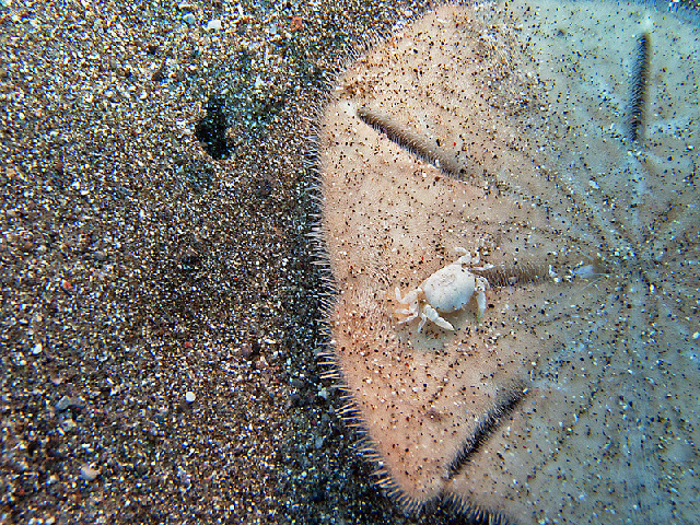
Adults of the Mediterranean Plumefoot Mite are considerably large for a mite, measuring a few millimeters in length, often more than 1 cm when the legs are considered. They are reddish-brown, lighter at the legs and other appendices, and their hind legs are much longer than the others and have a tuft of long black hair that makes them look like plumes, hence the name plumefoot mite. As the common name also suggest, this species is found around the Mediterranean, including southern Europe, northern Africa, Turkey and the Middle East.

Despite being a large and rather beautiful mite, very little is known about the life history of the Mediterranean Plumefoot Mite. It belongs to a group of mites that are predators as adults but parasites as larvae. The larvae hatch from red eggs laid by the female in the environment and are, of course, much smaller than the adults. They also have only three pairs of legs, and not four like the adults, and lack the characteristic plumes seen in the adults.

Some eggs of the Mediterranean Plumefoot Mite and one recently hatched larva. Extracted from Mąkol & Sevsay (2015).

Little to nothing is known about the feeding habits of this species. Grasshoppers are among the identified hosts of the larvae, but it is likely that other arthropods are parasitized as well. The larvae attach to the legs of the hosts and feed there, sucking their hemolymph (the “blood” of arthropods). I could not find any information about which species serve as prey for the adults.

Even though we know almost nothing about the ecology of the Mediterranean plumefoot mite, we can still appreciate its beauty, and it certainly plays a fundamental role in its ecosystem.

If you live around the Mediterranean, have you ever seen one of them? Let us know!

– – –

More mites:

Friday Fellow: Giant red velvet mite (on 22 July 2016)

Friday Fellow: Cuban-laurel-thrips mite (on 28 June 2019)

Friday Fellow: Rhinoceros Tick (on 18 October 2019)

Friday Fellow: Aloe Mite (on 7 February 2020)

– – –

Follow us on Twitter!

– – –

References:

GBIF. Eatoniana plumipes (L. Koch, 1856). Available at < https://www.gbif.org/species/4539982 >. Access on 20 October 2022.

Mąkol, J., & Sevsay, S. (2015). Abalakeus Southcott, 1994 is a junior synonym of “plume-footed” Eatoniana Cambridge, 1898 (Trombidiformes, Erythraeidae)-evidence from experimental rearing. Zootaxa3918(1), 92-112. https://doi.org/10.11646/zootaxa.3918.1.4

Noei, J., & Rabieh, M. M. (2019). New data on Nothrotrombidium, Southcottella and Eatoniana larvae (Acari: Trombellidae, Neothrombiidae, Erythraeidae) from Iran. Persian Journal of Acarology8(3). https://doi.org/10.22073/pja.v8i3.46776

– – –

*Creative Commons License This work is licensed under a Creative Commons Attribution-NonCommercial 4.0 International License.

Leave a comment

Filed under Arachnids, Friday Fellow, Parasites, Zoology

Friday Fellow: Deadly Plasmodium

by Piter Kehoma Boll

Humans can be infected by a vast number of different parasites, but no parasite has such a big impact on our species as those of the genus Plasmodium, which cause the disease called malaria. Several different species of Plasmodium infect humans, but today we will talk about the deadliest of them, Plasmodium falciparum, which I decided to name the Deadly Plasmodium.

The genus Plasmodium belongs to the phylum Apicomplexa, a group of exclusively parasitic protists that evolved from free-living algae. Besides Plasmodium, another important apicomplexan that infects humans is the Toxo, which causes toxoplasmosis and already appeared as a fellow here some years ago.

The life cycle of the deadly plasmodium is very complex and includes two hosts, a human and a mosquito of the genus Anopheles. When an infected mosquito bites a human, it releases between 20 and 200 sporozoites, which are one of the life stages of the deadly plasmodium. The sporozoites are elongated cells that glide through the bloodstream of the infected human until they reach the liver. They use their apical complex, a structure formed by several glands and organelles, to penetrate the liver cells. The apical complex is lost in the process.

Life cycle of the deadly plasmodium. Credits to La Roche Lab, UC Riverside.**

Inside the liver cells, the sporozoite changes into a trophozoite, which lives inside a vacuole and starts to grow and undergo mitosis and meiosis without cell division until becoming a single cell with tens of hundreds of nuclei called a schizont. The schizont eventually bursts into tens of thousands of small haploid cells called merozoites, which enter the bloodstream again. Provided with new apical complexes, the merozoites now use it to enter red blood cells and turn again into trophozoites, which are now haploid. This haploid red-cell-infecting trophozoite grows again and undergoes mitosis to make a new large schizont, which eventually bursts again into new merozoites that enter the bloodstream once more and infect new red cells, starting the cycle again. Inside the red cells, the trophozoite feeds on hemoglobin and converts part of it into an insoluble granular pigment called hemozoin.

A trophozoite (left) and a schizont (lower center) in red blood cells.

All plasmodium cells infecting the red blood cells have their cycle in synchrony so that all merozoites are released into the bloodstream and infect new cells simultaneously. The cycle in the red blood cells takes about 48 hours and is related to the typical cyclic manifestations of fever and chills caused by falciparum malaria.

Eventually, some merozoites change into sexual forms, the male and female gametocytes, known as microgametocyte and macrogametocyte, respectively. They take between one and two weeks to reach maturity. After reaching this stage, if the infected human is bitten by a mosquito, the gametocytes are ingested and, inside the mosquito’s gut, undergo morphological changes. The macrogametocytes grow into a large, spherical form, the macrogamete. The microgametocyte undergoes nuclear division in 15 minutes and splits into eight flagellated microgametes. A microgamete fertilizes a macrogamete, which originates a zygote. The zygote develops into a motile cell called the ookinete, which penetrates the mosquito tissues and turns into an immotile oocyst. The oocyst grows and undergoes nuclear division until becoming a large multinucleate cell called sporoblast. The sporoblast bursts into thousands of sporozoites, which migrate to the mosquito’s salivary glands and, when the mosquito bites another human, they are released into the bloodstream and restart the cycle.

A macrogametocyte (left) and a microgametocyte (right) among red blood cells.

The main deleterious effects caused by malaria in humans occur due to the massive destruction of red blood cells and the production of hemozoin, which is toxic to tissue and starts to accumulate in several organs, impairing their function. Although several species of Plasmodium can cause malaria in humans, about 50% of all deaths by malaria are caused by the deadly plasmodium alone, making it the deadliest parasite in humans.

The deadly plasmodium apparently evolved about 10 thousand years ago in Africa from a very similar species that infects gorillas. Currently, although most infections and deaths still occur in Subsaharan Africa, the deadly plasmodium has spread around the world and is also prevalent in regions near the equator in South America and Asia.

Due to the high lethality of falciparum malaria in humans, the disease has placed great selective pressure on human populations in areas where this parasite is common. Thus, although some say that natural selection does not affect humans anymore, the deadly plasmodium is here to refute this argument.

– – –

Other Apicomplexans:

Friday Fellow: Toxo (on 5 May 2017)

Friday Fellow: Arrow Anchor (on 15 January 2021)

– – –

Follow us on Twitter!

– – –

References:

Lambros, C., & Vanderberg, J. P. (1979). Synchronization of Plasmodium falciparum erythrocytic stages in culture. The Journal of parasitology, 418-420.

Maier, A. G., Matuschewski, K., Zhang, M., & Rug, M. (2019). Plasmodium falciparum. Trends in parasitology35(6), 481-482. https://doi.org/10.1016/j.pt.2018.11.010

Talman, A. M., Domarle, O., McKenzie, F. E., Ariey, F., & Robert, V. (2004). Gametocytogenesis: the puberty of Plasmodium falciparum. Malaria journal3(1), 1-14. https://doi.org/10.1186/1475-2875-3-24

Wikipedia. Plasmodium falciparum. Available at < https://en.wikipedia.org/wiki/Plasmodium_falciparum >.

– – –

**Creative Commons License This work is licensed under a Creative Commons Attribution 3.0 Unported License.

Leave a comment

Filed under Friday Fellow, Parasites, protists

Friday Fellow: Fungus Root

by Piter Kehoma Boll

Plants are the standard organism when one thinks of photosynthesis, but several species have actually lost the ability to synthesize their own food using light and have become completely heterotrophic. As a result, such plants survive by parasitizing other plants and feeding on their sap. Probably the most famous species of heterotrophic plant is Rafflesia arnoldii, the corpse flower, which has the largest flowers of any plant and was one one our first Friday Fellows almost 10 years ago. But today I will introduce you another, completely unrelated heterotrophic plant, Balanophora fungosa, commonly known as the fungus root.

This species occurs across southeast Asia and Australia, where it lives on the soil and parasitizes the roots of several different plants. As with most heterotrophic plants, the fungus root spends most of its life underground as nothing but a system of roots and rhizomes attached to the host plant. It is only visible on the surface when it produces its flowers, which, like the giant flower of the corpse flower, are also very unusual.

A group of inflorescences coming out of the ground in New Caledonia. We can see the pale bracts and the velvet-like club of female flowers surrounded by the larger male flowers at the base. Photo by iNaturalist user juju98.*

The flowers occur in inflorescences that are actually kind of cute. The overall color varies from pale cream, almost white, to pink. The base of the inflorescence has several bracts (modified, flower-associated leaves) that have the same pale cream to pink color, without any sign of green. The upper part has a club-shaped structure with a velvet-like surface formed by hundreds of tiny female flowers. Surrounding the base of the club are a few male flowers, which are much larger than the female flower, but still very small. The inflorescence as a whole looks similar to some mushrooms, such as puffballs, which is probably the reason for its common name fungus root.

A closeup of an inflorescence in Australia where we can see the male and female flowers in more details. Photo by Aaron Bean.*

I couldn’t find many details about the life cycle of this cute parasite, but it seems to be pollinated by an enormous range of animals, including several types of insects, arachnids and even small vertebrates, which may be attracted to feed on the pollen and nectar or perhaps tricked by the unusual smell that the flowers produce. The smell is unlike the sweet fragrance of most flowers but is also not an unpleasant smell of carrion like that of the corpse flower, the titan arum and so many other plants. Actually, it is said that the flowers smell like a mouse. Perhaps it tricks small mammals to think it is a reproductive member of their species just like some orchids do by mimicking the shape and smell of female bees? This is a possibility, but actually most of the small mammals and birds that visit the flowers are actually nectarivores and are probably only looking for the delicious nectar.

Anyway, there is a lot we still don’t know about this unusual but adorable fungus-like plant.

– – –

Follow us on Twitter!

– – –

References:

Hsiao, S. C., Huang, W. T., & Lin, M. S. (2010). Genetic diversity of Balanophora fungosa and its conservation in Taiwan. Botanical studies51(2). https://ejournal.sinica.edu.tw/bbas/content/2010/2/Bot512-10.pdf

Pierce, R., & Ogle, C. (2017). Musky Rat Kangaroos and other vertebrates feeding from the flowers of the root parasite’Balanophora fungosa’. North Queensland Naturalist47, 14-20. https://search.informit.org/doi/pdf/10.3316/informit.461078578600745

Wikipedia. Balanophora fungosa. Available at < https://en.wikipedia.org/wiki/Balanophora_fungosa >. Access on 8 July 2022.

– – –

*Creative Commons License This work is licensed under a Creative Commons Attribution-NonCommercial 4.0 International License.

Leave a comment

Filed under Botany, Parasites

Friday Fellow: Soybean cyst nematode

by Piter Kehoma Boll

In English, nematodes are known as roundworms, not because they are round like a ball, but because their body is cylindrical like a tube. However, some roundworms are indeed round, or sort of… These include the cyst nematodes of the genus Heterodera, one of which is Heterodera glycines, known as the soybean cyst nematode.

As you may guess by this species having “soybean” in its name, it is a parasite of soybeans. It is, indeed, one of the most devastating pests to attack soybean crops. These worms spend most of their life inside soybean roots, feeding on their tissues.

The typical aspect of the cysts on the roots of an infected plant.

Adults usually measure less than 1 mm in length but males and females are quite different in shape and behavior. Males are longer than females but are very slender, having the typical nematode shape. Once reaching adulthood, they leave the soybean roots and look for females to mate with. Females, on the other hand, have a lemon-shaped, almost spherical body. They never leave the soybean roots but, as they become sexually mature, their bodies swell with eggs, and eventually their posterior end bursts out of the roots, appearing as small white cysts visible to the naked eye.

An egg-filled female. Credits to Agroscope FAL Reckenholz , Swiss Federal Research Station for Agroecology and Agriculture.*

Filled with 200 to 400 eggs, the female dies and turns into a dark cyst. The eggs remain inside her dead body until the environmental conditions are adequate. During this period, the embryo develops from a Juvenile 1 (J1) into a Juvenile 2 stage (J2). The J2 leaves the egg and looks for a new soybean root to penetrate, where it then continues to develop until reaching adulthood.

A scanning electron micrograph showing an egg and a juvenile of the soybean cyst nematode.

The first symptoms of infection by the plant are very subtle, like a mild yellowing of the leaves, which is often mistaken for some nutrient deficiency. The problem is often only noticed during harvest, when yield losses can reach up to 30%.

There are still no effective ways to eliminate the soybean cyst nematode from infected areas. Some parasitic fungi are known to infect the nematode and control its population, and crop rotation is another strategy to reduce the infection load, as planting a different crop for two consecutive years shows a significant reduction in the number of viable eggs in the soil.

Life cycle of the soybean cyst nematode. Credits to George N. Agrios. Extracted from https://www.sciencedirect.com/topics/agricultural-and-biological-sciences/soybean-cyst-nematode

While there are some soybean varieties that are resistant to the infection, there are also different races of the soybean cyst nematode that show different degrees of virulence and new, often more virulent, races are constantly appearing to bypass the resistance developed by the plants. It is evolution being driven right in our farms.

– – –

Follow us on Twitter!

– – –

References:

Texas Invasive Species Institute. Soybean Cyst Nematode. Available at < http://www.tsusinvasives.org/home/database/heterodera-glycines >. Access on 30 June 2022.

Wikipedia. Soybean cyst nematode. Available at < https://en.wikipedia.org/wiki/Soybean_cyst_nematode >. Access on 30 June 2022.

Yan, G., & Baidoo, R. (2018). Current research status of Heterodera glycines resistance and its implication on soybean breeding. Engineering4(4), 534-541. https://doi.org/10.1016/j.eng.2018.07.009

– – –

*Creative Commons License This work is licensed under a Creative Commons Attribution-NonCommercial 4.0 International License.

Leave a comment

Filed under Parasites, worms

Friday Fellow: Human Chlamydia

by Piter Kehoma Boll

It’s time for another bacterium and why not one that loves us even though we hate it? Chlamydia trichomatis is today’s fellow, that little annoying bacterium that infects us humans and sometimes can cause us some serious problems.

The human chlamydia belongs to a phylum of bacteria known as Chlamydiae, which is simply the plural of the bacterium’s name. All Chlamydiae seem to be obligate endosymbionts of eukaryotic cells, either as parasites or in a mutualistic relationship. The human chlamydia, of course, is of the first type. This species is an exclusive parasite of humans and apparently cannot infect the cells of any other species.

The life cycle of the human chlamydia is similar to that of other chlamydia species. It has two distinct forms known as elementary bodies and reticulate bodies. The elementary bodies are a spore-like form measuring from 200 to 400 nanometers in diameter. They have a very rigid cell wall and are able to survive outside of a host cell. When an elementary body contacts a human host cell, mostly cells from the mucous membranes, it causes the host cell to make a vacuole in which it remains. This vacuole is known as an inclusion.

Chlamydia inclusions (the large bubbles in the central cell) as seen under the microscope.

Within the inclusion, the elementary body changes into the metabolically active reticulate body, which measures between 600 and 1500 nanometers. The reticulate body is capable to change the inclusion into a more suitable environment and starts to replicate very rapidly until filling the host cell with bacteria in up to 72 hours. At this point, the reticulate bodies change back to elementary bodies and make the host cell burst and release them, where they can spread to other cells and infect them as well.

The human chlamydia can infect many parts of the human body, but the most commonly affected areas are the urethra and the vagina and its transmission between humans occurs mainly through sexual intercourse as the infected person can have elementary bodies in its fluids, such as sperm and vaginal fluid. In fact, chlamydia is the most common sexually transmitted infection worldwide, with about 4.2% of all women and about 2.7% of all men having it.

Many cases of chlamydia infection can go undiagnosed because sometimes the infection does not cause any symptoms or they take a long time to appear. When it infects the vagina and cervix, symptoms are rare at first but as the infection spreads it can infect the rest of the reproductive system and cause pelvic inflammatory disease, which may lead to sterility. Some of the rare symptoms in a vaginal infection are pain during intercourse and vaginal bleeding. In the urethra, symptoms are more common and include pain or a burning sensation during urination and eventually an unusual discharge from the penis. The symptoms are very similar to those of gonorrhea.

Besides the urogenital tract, the human chlamydia can infect many other sites, such as the rectum and the oral cavity through anal and oral sex, respectively. Another commonly infected area are the eyes, with 19% of all cases of conjunctivitis being caused by the human chlamydia. If not adequately treated, this conjunctivitis evolves into a chronic condition known as trachoma that often leads to blindness. The eyes become infected by direct contact of infected hands or objects (such as towels) with the eyes. The bacterium can also be transmitted by flies as they move around human bodies licking their fluids.

Chlamydia infection by age and sex in the United States.

Chlamydia is often treated by antibiotics such as azithromycin. Prevention includes adequate hygiene, safe sexual practices and regular testing in sexually active humans since identifying the infection earlier can reduce its damage and prevent its spread to others. In the past decade, there has been an increased interest in developing a vaccine against chlamydia. One problem is the fact that the immune response against this bacterium seems to be very complex. However, preliminary tests with a candidate vaccine has led to promising results, so there is hope!

Despite being a pain in the ass (or most commonly in the crotch) for humans, the human chlamydia is at the same time a fascinating organism just like every other lifeform on Earth. It has a considerably small genome, with only bout 900 genes. Many essential metabolic genes are lacking and it is believed that they are scavenged from the host.

A human-specific parasite, the human chlamydia is believed to have become a separate lineage from other chlamydia species about 9 million years ago. This means it has been with us since before we were even humans.

– – –

Follow us on Twitter!

– – –

References:

Brunham, R. C., & Rey-Ladino, J. (2005). Immunology of Chlamydia infection: implications for a Chlamydia trachomatis vaccine. Nature reviews immunology5(2), 149-161. https://doi.org/10.1038/nri1551

Manavi, K. (2006). A review on infection with Chlamydia trachomatis. Best Practice & Research Clinical Obstetrics & Gynaecology20(6), 941-951. https://doi.org/10.1016/j.bpobgyn.2006.06.003

Wikipedia. Chlamydia trichomatis. Available at < https://en.wikipedia.org/wiki/Chlamydia_trachomatis >. Access on 13 January 2022.

– – –

1 Comment

Filed under Bacteria, Friday Fellow, Parasites

Friday Fellow: Pacific Waminoa

by Piter Kehoma Boll

A long time ago I talked about a polemic group of bilaterian animals, the acoelomorphs, whose exact phylogenetic position is uncertain but the evidence is starting to build up toward the idea that they are part of the first branch of bilaterians to diverge from the others, so that they are neither protostome nor deuterostomes. No acoelomorph has been presented as a Friday Fellow before, so today it is time for their debut. The species I selected is a very interesting one. Its name is Waminoa litus and, of course, it lacks a common name, so I decided to call it the Pacific waminoa because it is found in the Pacific, especially around Australia.

A marine species, the Pacific waminoa has the typical anatomy of the acoels, the subgroup of acoelomorphs to which it belongs. It lacks, for example, a gut, and its mouth opens directly into the internal tissues. The mouth is located at the venter, like in many flatworms, such as planarians. In fact, acoelomorphs were for a long time thought to be flatworms until molecular analyses revealed that they are not.

If you ever find small golden flakes on the surface of corals in Eastern Australia and nearby regions, you are most likely seeing several Pacific waminoa. Photo by Anne Hoggett.*

With a flat and circular, slightly heart-shaped body, the Pacific waminoa lives on the surface of corals. They are, in fact, kind of an ectoparasite to corals, as they feed on the coral’s mucus. But more than that, the Pacific waminoa also harbors symbiotic dinoflagellates (zooxanthellae) living directly inside its parenchyma, the tissue that fills their body. Zooxanthellae are famous as common symbiotic organisms living inside corals, and they share part of the nutrients they make via photosynthesis with their host. The corals in which the Pacific waminoa lives also harbor zooxanthellae. However, while all zooxanthellae living in the corals belong to the genus Symbiodinium, the ones living in the Pacific waminoa belong to two genera, Symbiodinium and Amphidinium. This is very unusual, as zooxanthellae belonging to two different genera in the same animal is a very rare occurrence. The beautiful golden color of the Pacific waminoa is a direct result of the presence of the zooxanthellae.

Several waminoas, likely Waminoa litus, on a coral in the Philippines. Photo by Franca Wermuth.*

At first it was thought that, due to this close relationship between these acoels and the corals, the lineages of Symbiodinium living inside both organisms would be closely related, but this is not the case. Molecular studies revealed that Symbiodinium living in the Pacific waminoa are not the same that live inside the corals, which indicates that both organisms acquired them independently, i.e., the Pacific waminoa does not captures them from the coral. In fact, they are passed directly from the mother to the offspring inside the eggs. The Pacific waminoa is born with the dinoflagellates already inside it.

The Pacific waminoa is, therefore, an incredible creature not only because it belongs to a very divergent lineage of bilaterian animals but because it is the only case known in bilaterians about symbionts being transferred directly from the mother to the offspring. Perhaps one day the zooxanthellae will evolve into a new organelle of a clade that will evolve from this amazing species.

– – –

Like us on Facebook!

Follow us on Twitter!

– – –

References:

Barneah, O., Brickner, I., Hooge, M., Weis, V. M., LaJeunesse, T. C., & Benayahu, Y. (2007). Three party symbiosis: acoelomorph worms, corals and unicellular algal symbionts in Eilat (Red Sea). Marine Biology151(4), 1215-1223.

Hikosaka-Katayama, T., Koike, K., Yamashita, H., Hikosaka, A., & Koike, K. (2012). Mechanisms of maternal inheritance of dinoflagellate symbionts in the acoelomorph worm Waminoa litus. Zoological science29(9), 559-567.

Kunihiro, S., & Reimer, J. D. (2018). Phylogenetic analyses of Symbiodinium isolated from Waminoa and their anthozoan hosts in the Ryukyu Archipelago, southern Japan. Symbiosis76(3), 253-264.

– – –

*Creative Commons License This work is licensed under a Creative Commons Attribution-NonCommercial 4.0 International License.

Leave a comment

Filed under Friday Fellow, Parasites

Friday Fellow: Fly-Killing Fungus

by Piter Kehoma Boll

You most likely know about the existence of the parasitic fungi of the family Cordycipitaceae that turn insects and other arthropods into “zombies”. I even presented one here already about 5 years ago, the Chinese caterpillar fungus. But zombie-making fungi did not evolve only once and today we will meet another one that also zombifies insects, more precisely flies.

Its scientific name is Entomophthora muscae and I decided to give it the common name fly-killing fungus. While the most famous zombifying fungi belong to the phylum Ascomycota, the fly-killing fungus belongs to the phylum Entomophtoromycota, which until recently was part of the phylum Zygomycota.

Dead fly of the species Scathophaga stercoraria consumed by the fly-killing fungus. Photo by Hans Hillewaert.*

The fly-killing fungus is actually a complex of species that infects a variety of flies, including house flies. Their life start as an asexual spore, the conidium. When the conidium contacts the surface of a fly, it germinates, forming a germ tube that penetrates the cuticle of the fly. When the germ tube reaches the haemocoel, the cavity in which the fly’s haemolymph (blood) is found, it releases some cells (protoplasts) into the haemolymph. The protoplasts are carried to the brain and grow in areas that control the fly’s behavior. From there, the fungus start to produce hyphae that grow through the whole body, slowly digesting the internal organs. When the fly is about to die, it crawls to a high point in the vegetation or other available structure, stretches the hind legs and opens the wings and eventually dies. Some hours later, the fungus start to produce conidiophores that grow on the surface of the fly. When they are mature, they release conidia into the environment, restarting the cycle.

Dead hoverfly of the species Melanostoma scalare with the conidiophores of the fly-killing fungus coming out of its abdomen. Photo by Wikimedia user TristramBrelstaff.**

The species complex known as Entomophthora muscae can infect a huge variety of flies, including domestic flies, fruit flies, blow flies, mosquitos and many others. As many of the hosts of the fly-killing fungus as medically or economically important species for humans, there have been attempts to “weaponize” this fungus, turning it into a biological control against several fly pests.

This is not an easy task, though. First, the fungus is very sensible to high temperatures and flies can “cure” themselves from the infection by moving to hot environments that inhibit the growth of the fungus. Another problem is that, in order to raise the fungus in the lab, the fungus needs a constant supply of live adults flies.

As in many cases of using biological agents to control pests, there is always the possibility of things going wrong and end up affecting species that were not the original target. The genetic diversity of the fly-killing fungus is not very well known yet, so that we don’t know exactly which fly species each strain (or species) can actually infect.

I hope we don’t end up turning this ecologically innocent fungus into one more villain on the planet due to our misbehavior.

– – –

Like us on Facebook!

Follow us on Twitter!

– – –

References and further reading:

Brobyn, P. J., & Wilding, N. (1983). Invasive and developmental processes of Entomophthora muscae infecting houseflies (Musca domestica). Transactions of the British Mycological Society80(1), 1-8. https://doi.org/10.1016/S0007-1536(83)80157-0

Kramer, J. P., & Steinkraus, D. C. (1981). Culture of Entomophthora muscae in vivo and its infectivity for six species of muscoid flies. Mycopathologia76(3), 139-143. https://doi.org/10.1007/BF00437194

Krasnoff, S. B., Watson, D. W., Gibson, D. M., & Kwan, E. C. (1995). Behavioral effects of the entomopathogenic fungus, Entomophthora muscae on its host Musca domestica: Postural changes in dying hosts and gated pattern of mortality. Journal of Insect Physiology41(10), 895-903. https://doi.org/10.1016/0022-1910(95)00026-Q

Wikipedia. Enthomophthora muscae. Available at < https://en.wikipedia.org/wiki/Entomophthora_muscae >. Access on 12 August 2021.

– – –

*Creative Commons License This work is licensed under a Creative Commons Attribution-ShareAlike 4.0 International License.

**Creative Commons License This work is licensed under a Creative Commons Attribution-ShareAlike 3.0 Unported License.

Leave a comment

Filed under Friday Fellow, Fungi, Parasites

Friday Fellow: Coldwater Disease Bacterium

by Piter Kehoma Boll

Bacteria are some of the most diverse organisms on Earth and they perform all sort of ecological roles, although they are more often associated with diseases by the average human being. This is, of course, due to the fact that most bacteria that have a direct and perceptible influence on human life are, in fact, pathogenic, often parasitic, bacteria. Today I am introducing one of those pathogenic bacteria, but not one that infects humans.

Currently known as Flavobacterium psychrophilum, this species is known to infect freshwater fish, causing a disease known as bacterial coldwater disease (CWD). As a result, I will call this species the coldwater disease bacterium, or CWD bacterium for short.

The typical aspect of CWD bacteria seen under the microscope. Extracted from Cipriano & Holt (2005).

The CWD bacterium is a typical rod-shaped bacterium measuring up to 1 µm in width and 5 µm in length. It lacks any type of flagellum or other structure that helps it move, but it can move by gliding, although this movement is incredibly slow and very difficult to observe. When cultured in a growth medium, they produce small 3-mm-diameter yellow colonies with thin margins.

Several colonies of CDW bacteria on a culture medium. Credits to Eva Säker (SVA) & Karl-Erik Johansson (BVF, SLU & SVA).*

Living in freshwater, the CWD bacterium prefers cold waters, with temperatures of 16 °C or lower, with the optimal temperature being 13°C. They grow on all sort of tissues on the body of fish, such as the skin, gills, mucous and internal organs such as the brain, kidney, spleen and the sex organs. Its preferred hosts are salmonoid fishes, such as salmons and trouts, but it can be found in other species eventually.

The CWD bacterium is an aerobic bacterium but is unable to use carbohydrates as a source of carbon, feeding on peptides instead. Thus, it secretes enzymes on the host’s tissues to degrade its proteins into peptides, causing structural damage.

Infected fish show tissue erosion, which often begins on the caudal fin and eventually spread. Fins become dark, ragged, split or torn and may be completely lost. Ulcerations appear on the skin, especially around the jaw, and the fish present behavioral issues such as spiral swimming and lethargy. The infection often kills the fish but sometimes a milder chronic infection can occur, which, however, still causes considerable behavioral changes in the host.

Lesions caused by Flavobacterium psychrophilum in the rainbow trout Oncorhynchus mykiss (A) and the coho salmon Oncorhynchus kisutsch. Extracted from Starliper et al. (2011).

The bacteria are often transmitted from fish to fish via direct fish contact, but infected adult fish can end up passing the infection directly to their offspring through infected eggs. The infection can be treated in early stages using the antibiotic oxytetracline or by adding quaternary ammonium cations to the water.

In natural environments the problems caused by this infection are rarely problematic and its damage is more often seen in fish farms, where the poor creatures are forced to live in higher densities, which increases the bacterium’s success. Apparently native to North America, where it was discovered in the 1940s, it was spread via fish farming across the whole world in the following decades. We humans, therefore, are once more the main reason why this species has become a worldwide problem.

– – –

References:

Cipriano, R. C., & Holt, R. A. (2005). Flavobacterium psychrophilum, cause of bacterial cold-water disease and rainbow trout fry syndrome. Kearneysville, WV: US Department of the Interior, US Geological Survey, National Fish Health Research Laboratory.

Langevin, C., Blanco, M., Martin, S. A., Jouneau, L., Bernardet, J. F., Houel, A., … & Boudinot, P. (2012). Transcriptional responses of resistant and susceptible fish clones to the bacterial pathogen Flavobacterium psychrophilum. PLoS One7(6), e39126. https://doi.org/10.1371/journal.pone.0039126

Starliper, C. E. (2011). Bacterial coldwater disease of fishes caused by Flavobacterium psychrophilum. Journal of Advanced Research2(2), 97-108. https://doi.org/10.1016/j.jare.2010.04.001

Wikipedia. Flavobacterium psychrophilum. Available at < https://en.wikipedia.org/wiki/Flavobacterium_psychrophilum >. Access on 10 June 2020.

– – –

*Creative Commons License This work is licensed under a Creative Commons Attribution 2.5 Sweden Generic License.

1 Comment

Filed under Bacteria, Friday Fellow, Parasites

Friday Fellow: Sand-Dollar Pea Crab

by Piter Kehoma Boll

Last week I presented the lovely five-slotted sand dollar, a very common echinoderm along the Atlantic Coast of the Americas. But as we all know, species rarely live by themselves. All kinds of association exist between organism, and today our species is one that lives closely associated with the five-slotted sand dollar, the sand-dollar pea crab, Dissodactylus mellitae.

The sand-dollar pea crab is, like its name suggests, a crab. A very very small crab indeed. Adult males reach up to 3.5 mm in size and females do not grow larger than 4.5 mm. They have a light-yellow to white color, sometimes with a complex pattern of darker marks on the dorsum.

The tiny sand-dollar pea crab. Credits to Naturalist Biodiversity Center.

The natural habitat of the sand-dollar pea crab is the surface of sand dollars, especially the five-slotted sand dollar. As they are very small, they live very comfortably among the hairs and spines of their host, most commonly on their ventral side, protected from light and possible predators.

For some time it was unknown whether the relationship between both species was that of commensalism, where the crab only eats together with the sand dollar, or of parasitism, where the crabs steals food from the sand dollar or feeds on the sand dollar itself. Analysis of the stomach content of the crabs revealed that up to 80% of its diet consists of tissues of the sand dollar, so that their relationship is most likely parasitic. In fact, it has been shown that the presence of the crabs reduces the number of eggs that female sand dollars produce.

Sand-dollar pea crab on its host. Credits to Naturalist Biodiversity Center.*

The maximum number of crabs observed on a single sand dollar was 10, but this number is highly dependent on the crab’s life stage. In summer, juveniles often prefer to live together, sharing the same host, but as they grow they disperse and prefer a solitary life, not sharing their host with others of the same species. When they are sexually mature, though, they often share the host with another crab of the opposite sex, thus facilitating reproduction. However, males seem to be much more common in the population, so males pairing with other males are more common than males pairing with females.

Reproduction seems to occur around late summer and fall along the coast of North America, after which the number of adult crabs starts to decrease. Juveniles start to appear in late spring, eagerly looking for sand dollars to colonize.

In the sea, different species associate even more frequently than on land. And we know that wherever there is life, there will be another life to parasitize it.

– – –

Like us on Facebook!

Follow us on Twitter!

– – –

References:

George, S. B., & Boone, S. (2003). The ectosymbiont crab Dissodactylus mellitae–sand dollar Mellita isometra relationship. Journal of Experimental Marine Biology and Ecology294(2), 235-255. https://doi.org/10.1016/S0022-0981(03)00271-5

Telford, M. (1982). Echinoderm spine structure, feeding and host relationships of four species of Dissodactylus (Brachyura: Pinnotheridae). Bulletin of Marine Science32(2), 584-594. https://www.ingentaconnect.com/content/umrsmas/bullmar/1982/00000032/00000002/art00017

– – –

*Creative Commons License This work is licensed under a Creative Commons Attribution-NonCommercial-Share Alike 3.0 Unported License.

Leave a comment

Filed under crustaceans, Friday Fellow, Parasites

Friday Fellow: Fire-Ant Internal Microsporidium

by Piter Kehoma Boll

Last week we learned about the red imported fire ant and the nuisance that it presents in North America, where it was introduced almost a century ago and has turned into a threat to ecosystems and people. As chemical pesticides are often harmful to species other than the one targeted, more species-specific alternatives are often sought. One of those is biological control, which can also be problematic if incorrectly used.

Today’s species is one of those biological agents that can help control the invasion of the red imported fire ant. Named Kneallhazia solenopsae, which I decided to nickname as the fire-ant internal microsporidium, this species belongs to the division Microsporidia, which are single-celled fungi. I already presented one of them here several years ago, the flounder glugea.

The fire-ant internal microsporidium is known to infect fire ants, including the red imported fire ant. It has a very complex life cycle with several stages that is not completely understood yet.

The most serious infection occurs in queens, in which the life stage known as type 2 dikaryotic spore (Type 2 DK) infects the fat body tissue. This is a stage of the microsporidium in which the cell has two nuclei, hence the name. Other adult ants are also infected similarly. The infection spreads slowly, causing hypertrophy of the fat body tissue and, at advanced infections stages, makes this tissue be completely replaced by masses of spores, preventing the development and reproduction of the queen. However, it takes a long time for the microsporidium to cause such a serious disease. It does not mean that it is doing nothing to make things worse before that, though.

While the fat body tissue of the queen is being slowly consumed by the Type 2 DK spores, many of them turn into another stage, the octospore, so called because it occurs in a vesicle which contains eight of them. These vesicles are relatively large compared to the other stages and are thought to be the infectious stage. An injured or dead ant, which is often cannibalized by others, releases the vesicles in the environment. They need to be ingested for infection to occur, but only 4-instar larvae are able to swallow them, because all other larval stages, as well as adults, have a “filter” in their mouth that prevents them ingesting particles larger than 0.8 µm and the vesicles are larger than that.

Several vesicles with octospores and a Type 2 DK spore of the fire-ant internal microsporidium. Photo by David Williams.**

Inside the 4-instar larvae, the vesicles release the octospores, which infect the larvae and fuse, turning into another type of cell with two nuclei, the Type 1 DK spores. Those spores are only found in 4-instar larvae and pupae and are supposed to turn into Type 2 DK spores during the development into adult ants, where they can produce new octospores.

But this is not the only way the microsporidium spreads through the colony. Going back to the queens, the Type 2 DK spores also infect their ovaries and, after the queen is inseminated, they turn into another stage, the so-called megaspore. These megaspores infect the eggs and are incorporated into the embryo and pass directly to the 1-instar larvae, where they develop into Type 1 DK spores as they grow.

So the only way for an ant to become infected is if it receives the parasite directly from the queen in the egg or if it ingests the octospore vesicles as a 4-instar larva? No. And the reason is the fact that ants perform trophallaxis, which is the transfer of food from one individual to the other, i.e., the ants vomit into each other’s mouth. Thus, if an adult ant exchanges food with a 4-instar larva that has released octospores in its gut, it may end up infected as well.

That is quite a complicated life cycle, right? But it seems to be quite well tuned for the microsporidium to be a successulf parasite of fire ants. Is it an efficient biological control though? Well, I would say that it may help slowing down the spread of fire ants, but as its effects causing sterility to the queen take a long time to appear, it will not lead to the removal of the ants in areas they have invaded already.

– – –

Like us on Facebook!

Follow us on Twitter!

– – –

References:

Knell, J. D., Allen, G. E., & Hazard, E. I. (1977). Light and electron microscope study of Thelohania solenopsae n. sp.(Microsporida: Protozoa) in the red imported fire ant, Solenopsis invicta. Journal of Invertebrate Pathology29(2), 192-200. https://doi.org/10.1016/0022-2011(77)90193-8

Oi, D. H., & Williams, D. F. (2002). Impact of Thelohania solenopsae (Microsporidia: Thelohaniidae) on polygyne colonies of red imported fire ants (Hymenoptera: Formicidae). Journal of Economic Entomology95(3), 558-562. https://doi.org/10.1603/0022-0493-95.3.558

Sokolova, Y. Y., & Fuxa, J. R. (2008). Biology and life-cycle of the microsporidium Kneallhazia solenopsae Knell Allan Hazard 1977 gen. n., comb. n., from the fire ant Solenopsis invictaParasitology135(8), 903.

– – –

**Creative Commons License This work is licensed under a Creative Commons Attribution 3.0 Unported License.

Leave a comment

Filed under Friday Fellow, Fungi, Parasites, protists