Monthly Archives: August 2021

Friday Fellow: Striped Barnacle

by Piter Kehoma Boll

Barnacles are a peculiar group of sessile crustaceans and are common worldwide in the oceans, sometimes even living on the surface of marine animals such as mollusks, whales and turtles. Until now only one barnacle was featured here, a goose barnacle. So today we will know a species of the most common acorn barnacles, Amphibalanus amphitrite, known as the striped barnacle, purple acorn barnacle or Amphitrite’s rock barnacle.

The purple vertical stripes give the striped barnacle its name. Photo by iNaturalist user julieskrinni.*

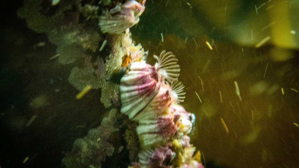
The striped barnacle has the typical conical shape of acorn barnacles formed by six calcareous plates surrounding the body. The opening at the top has a diamond shape and is protected by a movable lid formed by two plates. The plates of the shell have a series of vertical brown to purple stripes, hence the name striped barnacle. To eat, the striped barnacle opens the lid and extends its long feathery legs, called cirri, through the opening to capture food particles from the water.

Submerged striped barnacles with their cirri exposed. Photo by Jason Lee Boswell.*

The exact origin of the striped barnacle is unknown, but it is likely native from the Indian ocean. However, due to human activities, it has been carried across the whole world and is now found in warm and temperate waters of all oceans.

As typical of barnacles, the striped barnacle is hermaphrodite. To reproduce, they use a very long penis that they insert inside adjacent barnacles to release sperm. The fertilized eggs are released in the water and develop first into a nauplius larva os crustaceans and later into the cyprid larva, which is the last stage before they become adults. The cyprid looks for an adequate substrate to settle and, once finding it, starts to secrete a glycoproteinous substance to attach the head on the substrate and undergoes the final metamorphosis to became a juvenile barnacle. They continue to molt as they keep growing after attaching to the substrate, but the calcarous plates do not molt with them, but continue to grow like the shell of a mollusk.

Several striped barnacles growing on the back of a horseshoe crab. Photo by iNaturalist user ozarkpoppy.*

The striped barnacle can grow on human-made structures, such as ships, pipes and other constructions exposed to the tides, and can become a nuisance, as its presence can decrease the efficacy of some of the colonized structures. As a result, it has become a target species of studies and is even a model organism for the study of larval settlement of barnacles. Even its genome has already been sequenced and several technologies are being tested to reduce its ability to colonize ships.

Not all studies with this species are directed to ways to getting rid of it, though. Due to the ease of breeding it in the lab, the striped barnacle is also used to study, for example, the impact of microplastics and ocean acidification on marine life.

– – –

Like us on Facebook!

Follow us on Twitter!

– – –

References and further reading:

Bhargava, S., Chen Lee, S. S., Min Ying, L. S., Neo, M. L., Lay-Ming Teo, S., & Valiyaveettil, S. (2018). Fate of nanoplastics in marine larvae: a case study using barnacles, Amphibalanus amphitrite. ACS Sustainable chemistry & engineering6(5), 6932-6940. https://pubs.acs.org/doi/abs/10.1021/acssuschemeng.8b00766

Burden, D. K., Spillmann, C. M., Everett, R. K., Barlow, D. E., Orihuela, B., Deschamps, J. R., … & Wahl, K. J. (2014). Growth and development of the barnacle Amphibalanus amphitrite: time and spatially resolved structure and chemistry of the base plate. Biofouling30(7), 799-812. https://doi.org/10.1080/08927014.2014.930736

Maréchal, J. P., & Hellio, C. (2011). Antifouling activity against barnacle cypris larvae: Do target species matter (Amphibalanus amphitrite versus Semibalanus balanoides)?. International Biodeterioration & Biodegradation65(1), 92-101. https://doi.org/10.1016/j.ibiod.2010.10.002

McDonald, M. R., McClintock, J. B., Amsler, C. D., Rittschof, D., Angus, R. A., Orihuela, B., & Lutostanski, K. (2009). Effects of ocean acidification over the life history of the barnacle Amphibalanus amphitrite. Marine Ecology Progress Series385, 179-187. https://doi.org/10.3354/meps08099

– – –

Leave a comment

Filed under Conservation, crustaceans, Friday Fellow

Friday Fellow: Eelgrass Labyrinthula

by Piter Kehoma Boll

In the 1930s, a devastating disease spread across the Atlantic populations of the common eelgrass, killing 90% of the plants. The disease started by destructing the plant’s chloroplasts, turning the leaves white, followed by a rot of the tissues that spread until the whole plant was dead.

Although the agent causing the disease was already suggested back then, it was only confirmed during a second, smaller recurrence in the 1980s. The responsible is a marine fungi-like parasite known as Labyrinthula zosterae, which I decided to call the eelgrass labyrinthula.

Dark marks of rotten tissue in the common eelgrass caused by the eelgrass labyrinthula. Extracted from Short (2014).

The eelgrass labyrinthula belongs to a peculiar group of organisms that were at first classified as a weird group of slime molds but are now known to be heterokonts, thus more closely related to diatoms, brown algae and oomycetes, for example. They are colonial organisms and their colony is quite interesting in its arrangement. The individual vegetative cells are spindle-shaped (fusiform) and measure from 15 to 20 µm in length by 3 to 5 µm in width. They are often translucent, sometimes pale yellow, and are filled with lipid droplets.

Vegetative cells of the eelgrass labyrinthula. The ectoplasmic net can be seen as thin white extensions. Extracted from theredshrimp.com

The cells produce together a net formed by ectoplasm (cytoplasm) that is excreted from the cells and is surrounded by a cell membrane, thus becoming kind of like a large maze-like cell inside of which the individual cells live. This net lacks a cell wall and organelles, though. The cells can slide across this net, using it as a “road”.

There are two modes or reproduction known until now. The first is simply by cell division through mitosis. Another form of reproduction is by zoosporulation, which starts with several vegetative cells aggregating and fusing into a single plasmodium-like structure. Later, this plasmodium divides back into round, enlarged presporangia, which then divide internally into eight zoospores, which are then released in the environment. The zoospores are cells with two flagella and a stigma (eyespot), very similar to the typical cell of other heterokonts, and will differentiate back into vegetative cells.

The infection of the common eelgrass by the eelgrass labyrinthula occurs by direct contact of a healthy plant with an infected one or with dettached, infected parts. The parasite acts by dissolving the plant’s cell wall and spreading across the tissues, destroying the cells and likely feeding on them. Recent studies, however, discovered that the eelgrass labyrinthula is quite common in eelgrass populations but most strains are not very virulent. In fact, it is likely that the eelgrass labyrinthula is part of the “normal” protist biota associated with the common eelgrass and it only becomes a threat when the plant experience some sort of stress induced by other conditions.

Anyway, there is still much to be discovered about this protist and its dubious intentions.

– – –

Like us on Facebook!

Follow us on Twitter!

– – –

References:

Brakel, J., Werner, F. J., Tams, V., Reusch, T. B., & Bockelmann, A. C. (2014). Current European Labyrinthula zosterae are not virulent and modulate seagrass (Zostera marina) defense gene expression. PLoS One9(4), e92448. https://doi.org/10.1371/journal.pone.0092448

Muehlstein, L. K., Porter, D., & Short, F. T. (1991). Labyrinthula zosterae sp. nov., the causative agent of wasting disease of eelgrass, Zostera marina. Mycologia83(2), 180-191. https://doi.org/10.1080/00275514.1991.12025994

Ralph, P. J., & Short, F. T. (2002). Impact of the wasting disease pathogen, Labyrinthula zosterae, on the photobiology of eelgrass Zostera marinaMarine Ecology Progress Series226, 265-271.

Short, Fred, “Eelgrass Wasting Disease: an Overview” (2014). Salish Sea Ecosystem Conference. 34.
https://cedar.wwu.edu/ssec/2014ssec/Day1/34

Leave a comment

Filed under Friday Fellow, protists

Friday Fellow: Common Eelgrass

by Piter Kehoma Boll

Today we reach 300 Friday fellows, dear readers! And, as usual we will celebrate this presenting two species today.

Due to this special occasion, let’s present a special species as well, the common eelgrass, Zostera marina. But if it is special, why is it called common?

Well, this species is special because it is a marine flowering plant. Although flowering plants have conquered the planet across almost all its biomes, they are pretty rare in the sea, but the common eelgrass is one exception. And it is such a successful species that it can be found around the whole northern hemisphere in the Atlantic and Pacific oceans, including the Mediterranean and the Black Sea.

Common eelgrass at the northwest coast of the USA. Photo by John Brew.*

The ideal habitat for the common eelgrass are cold, clear and shallow waters, although not too shallow, as it likes to remain completely submerged. Its stem is a rhizome that grows inside the sandy or muddy substrate and from which long slender leaves emerge. The leaves have about 1 cm in width but their length can vary from a few centimeters to more than one meter depending on the water depth and turbidity.

The common eelgrass is able to reproduce entirely underwater. The flowers arise from the base of the leaves and form an elongate inflorescence with alternating female and male flowers that is enclosed in a sheath. Female flowers develop first, exposing their styles and waiting to be pollinated by the pollen of other plants. The pollen is specially adapted to be transported through the water. After the female flowers have been pollinated, their styles retract and the male flowers expose their anthers to release pollen in the water. The fruits start to develop and form a small transparent sack that contains the seed. After seed maturation, the fruits are released in the water to find a substrate to germinate.

Flowering stages of the common eelgrass. (1) female flowers expose their styles; (2) pollinated female flowers retract to start seed development; (3) male flowers expose the anthers and release pollen; (4) seeds starting to mature; (5) mature seeds ready to be released. Extracted from Infantes & Moksnes (2018).

Human populations have used the common eelgrass for centuries or even millenia. They can be harvested to be used as a fertilizer or fodder to cattle, the dried leaves can be used to stuff mattresses and they can also serve as food, with both the leaves and seeds being edible.

The stable environment formed by the common eelgrass is an important habitat for many marine species, including animals, algae and even bacteria, which use them as shelter, reproductive site or even feed on the plants. However, the increasing turbidity in coastal waters caused by human activities, especially water pollution, threatens many populations of this species, as it cannot adequately photosynthesize if light is blocked in the water column.

– – –

Like us on Facebook!

Follow us on Twitter!

– – –

References:

Infantes, E., & Moksnes, P. O. (2018). Eelgrass seed harvesting: Flowering shoots development and restoration on the Swedish west coastAquatic botany144, 9-19.

Wikipedia. Zostera marina. Available at < https://en.wikipedia.org/wiki/Zostera_marina >. Access on 19 August 2021.

– – –

*Creative Commons License This work is licensed under a Creative Commons Attribution 4.0 International License.

1 Comment

Filed under Botany, Friday Fellow

Friday Fellow: Fly-Killing Fungus

by Piter Kehoma Boll

You most likely know about the existence of the parasitic fungi of the family Cordycipitaceae that turn insects and other arthropods into “zombies”. I even presented one here already about 5 years ago, the Chinese caterpillar fungus. But zombie-making fungi did not evolve only once and today we will meet another one that also zombifies insects, more precisely flies.

Its scientific name is Entomophthora muscae and I decided to give it the common name fly-killing fungus. While the most famous zombifying fungi belong to the phylum Ascomycota, the fly-killing fungus belongs to the phylum Entomophtoromycota, which until recently was part of the phylum Zygomycota.

Dead fly of the species Scathophaga stercoraria consumed by the fly-killing fungus. Photo by Hans Hillewaert.*

The fly-killing fungus is actually a complex of species that infects a variety of flies, including house flies. Their life start as an asexual spore, the conidium. When the conidium contacts the surface of a fly, it germinates, forming a germ tube that penetrates the cuticle of the fly. When the germ tube reaches the haemocoel, the cavity in which the fly’s haemolymph (blood) is found, it releases some cells (protoplasts) into the haemolymph. The protoplasts are carried to the brain and grow in areas that control the fly’s behavior. From there, the fungus start to produce hyphae that grow through the whole body, slowly digesting the internal organs. When the fly is about to die, it crawls to a high point in the vegetation or other available structure, stretches the hind legs and opens the wings and eventually dies. Some hours later, the fungus start to produce conidiophores that grow on the surface of the fly. When they are mature, they release conidia into the environment, restarting the cycle.

Dead hoverfly of the species Melanostoma scalare with the conidiophores of the fly-killing fungus coming out of its abdomen. Photo by Wikimedia user TristramBrelstaff.**

The species complex known as Entomophthora muscae can infect a huge variety of flies, including domestic flies, fruit flies, blow flies, mosquitos and many others. As many of the hosts of the fly-killing fungus as medically or economically important species for humans, there have been attempts to “weaponize” this fungus, turning it into a biological control against several fly pests.

This is not an easy task, though. First, the fungus is very sensible to high temperatures and flies can “cure” themselves from the infection by moving to hot environments that inhibit the growth of the fungus. Another problem is that, in order to raise the fungus in the lab, the fungus needs a constant supply of live adults flies.

As in many cases of using biological agents to control pests, there is always the possibility of things going wrong and end up affecting species that were not the original target. The genetic diversity of the fly-killing fungus is not very well known yet, so that we don’t know exactly which fly species each strain (or species) can actually infect.

I hope we don’t end up turning this ecologically innocent fungus into one more villain on the planet due to our misbehavior.

– – –

Like us on Facebook!

Follow us on Twitter!

– – –

References and further reading:

Brobyn, P. J., & Wilding, N. (1983). Invasive and developmental processes of Entomophthora muscae infecting houseflies (Musca domestica). Transactions of the British Mycological Society80(1), 1-8. https://doi.org/10.1016/S0007-1536(83)80157-0

Kramer, J. P., & Steinkraus, D. C. (1981). Culture of Entomophthora muscae in vivo and its infectivity for six species of muscoid flies. Mycopathologia76(3), 139-143. https://doi.org/10.1007/BF00437194

Krasnoff, S. B., Watson, D. W., Gibson, D. M., & Kwan, E. C. (1995). Behavioral effects of the entomopathogenic fungus, Entomophthora muscae on its host Musca domestica: Postural changes in dying hosts and gated pattern of mortality. Journal of Insect Physiology41(10), 895-903. https://doi.org/10.1016/0022-1910(95)00026-Q

Wikipedia. Enthomophthora muscae. Available at < https://en.wikipedia.org/wiki/Entomophthora_muscae >. Access on 12 August 2021.

– – –

*Creative Commons License This work is licensed under a Creative Commons Attribution-ShareAlike 4.0 International License.

**Creative Commons License This work is licensed under a Creative Commons Attribution-ShareAlike 3.0 Unported License.

Leave a comment

Filed under Friday Fellow, Fungi, Parasites

Friday Fellow: Brazilian Rain Lily

by Piter Kehoma Boll

Today species is once again one that was very special to me during my childhood. Its scientific name is either Habranthus robustus or Zephyranthes robusta. I’m not sure which one is currently valid. Its common name in pink rain lily, Brazilian rain lily and many others. It is, however, not a true lily, but a species of the family Amaryllidaceae in the order Asparagales, thus more closely related to things like onions, asparagus and even orchids.

Like most species of Amaryllidaceae and other lily-like monocts, the Brazilian rain lily has an underground bulb that persists through the year. During summer, especially after rainy days, a long scape (stalk), up tp 30 cm long, develops from the bulb and will originate a single flower at its end.

Two beautiful flowers photographed in São Paulo. Photo by Luís Roberto Silva.**

The flowers are up to 6 cm long and have a lavender to pale pink color and a typical lilioid shape, with three petals and three petal-like sepals, as well as six stamens and a single pistil. A single bract (flower-associated leaf) can be seen at the base of the scape. The flowers do not last more than a couple of days and develop into a three-segmented fruit, which will eventually get dry and open to release the seeds.v

Fruits and buds side by side in Penha, Brazil. Photo by iNaturalist user luisfunez**.

Usually after flowering, the plant will produce a group of leaves. These are long, narrow, flat and smooth and are covered by a thin layer of a silverly waxy substance, which gaves them a slightly bluish tinge.

Here you can see the bluish tinge caused by the grayish wax on the leaves. Photo by Krzysztof Ziarnek.*

The Brazilian rain lily is native from southern Brazil and neighboring regions of Argentina, Uruguay and Paraguay but due to its beauty it has become a considerably popular ornamental species and has become naturalized in other parts of the world, especially South Africa and southern United States.

The plant is mildly toxic and can cause irritation in the mucous membranes if ingested. Some alkaloids extracted from the bulbs showed to have acetylcholinesterase- and butyrylcholinesterase-inhibitory activities, which indicates their potential for the development of new drugs for the treatment of dementia.

– – –

Like us on Facebook!

Follow us on Twitter!

– – –

References:

Kulhánková, A., Cahlíková, L., Novák, Z., Macáková, K., Kuneš, J., & Opletal, L. (2013). Alkaloids from Zephyranthes robusta Baker and their acetylcholinesterase‐and butyrylcholinesterase‐inhibitory activity. Chemistry & biodiversity10(6), 1120-1127. https://doi.org/10.1002/cbdv.201200144

Wikipedia. Habranthus robustus. Available at < https://en.wikipedia.org/wiki/Habranthus_robustus >. Access on 4 August 2021.

– – –

*Creative Commons License This work is licensed under a Creative Commons Attribution-ShareAlike 4.0 International License.

**Creative Commons License This work is licensed under a Creative Commons Attribution-NonCommercial 4.0 International License.

Leave a comment

Filed under Botany, Friday Fellow