Ostracods, or seed shrimps, are very small and very abundant crustaceans, but also very neglected by biologists. I had presented three species here before, including a bioluminescent one, and it is time for the next one. Today’s species is named Chlamydotheca spinosa and I decided to give it the common name Jamaican Seed Shrimp.
As the common name suggests, the Jamaican seed shrimp occurs in Jamaica, but it can also be found in several other Caribbean islands and in Central and South America. It is a freshwater seed shrimp and one of the largest freshwater seed shrimps with more than 6 mm in length. As in all seed shrimps, its body is covered by two shells. The shells are elongate and narrow, with a small point at the back, and bluish green marks on them.
A preserved specimen with faded colors. Extracted from Schmidt et al. (2018).*
Living at the bottom of freshwater ponds, the Jamaican seed shrimp is probably a deposit feeder, ingesting organic matter and smaller organisms, such as algae. It can be raised in aquaria very easily, which would make it a very good model for studies, only that no one seems to care. Another interesting thing about this species is the fact that it is parthenogenetic, which means females produce offspring from unfertilized eggs so that males do not exist.
Like all arthropods, the Jamaican seed shrimp molts as it grows. Its weight doubles at each molt, except for the last molt, between the eighth instar and the adult form, when it increases a little bit more, probably because of the development of the gonads.
And that’s all I could find about this little fellow. If you work with ostracods, try to provide more information about them online. That would be very useful to help us spread knowledge and love for these amazing animals.
Kesling, R. V., & Crafts, F. C. (1962). Ontogenetic increase in Archimedean weight of the ostracod Chlamydotheca unispinosa (Baird). American Midland Naturalist, 149-153. https://www.jstor.org/stable/2422641
Schmidt, R. E., Shoobs, N. F., & McMullin, E. R. (2018). Occurrence of the large ostracod, Chlamydotheca unispinosa (Baird, 1862), in temporary waters of Montserrat, Lesser Antilles. ZooKeys, (748), 89. https://doi.org/10.3897/zookeys.748.22323
It’s time for another bacterium and why not one that loves us even though we hate it? Chlamydia trichomatis is today’s fellow, that little annoying bacterium that infects us humans and sometimes can cause us some serious problems.
The human chlamydia belongs to a phylum of bacteria known as Chlamydiae, which is simply the plural of the bacterium’s name. All Chlamydiae seem to be obligate endosymbionts of eukaryotic cells, either as parasites or in a mutualistic relationship. The human chlamydia, of course, is of the first type. This species is an exclusive parasite of humans and apparently cannot infect the cells of any other species.
The life cycle of the human chlamydia is similar to that of other chlamydia species. It has two distinct forms known as elementary bodies and reticulate bodies. The elementary bodies are a spore-like form measuring from 200 to 400 nanometers in diameter. They have a very rigid cell wall and are able to survive outside of a host cell. When an elementary body contacts a human host cell, mostly cells from the mucous membranes, it causes the host cell to make a vacuole in which it remains. This vacuole is known as an inclusion.
Chlamydia inclusions (the large bubbles in the central cell) as seen under the microscope.
Within the inclusion, the elementary body changes into the metabolically active reticulate body, which measures between 600 and 1500 nanometers. The reticulate body is capable to change the inclusion into a more suitable environment and starts to replicate very rapidly until filling the host cell with bacteria in up to 72 hours. At this point, the reticulate bodies change back to elementary bodies and make the host cell burst and release them, where they can spread to other cells and infect them as well.
The human chlamydia can infect many parts of the human body, but the most commonly affected areas are the urethra and the vagina and its transmission between humans occurs mainly through sexual intercourse as the infected person can have elementary bodies in its fluids, such as sperm and vaginal fluid. In fact, chlamydia is the most common sexually transmitted infection worldwide, with about 4.2% of all women and about 2.7% of all men having it.
Many cases of chlamydia infection can go undiagnosed because sometimes the infection does not cause any symptoms or they take a long time to appear. When it infects the vagina and cervix, symptoms are rare at first but as the infection spreads it can infect the rest of the reproductive system and cause pelvic inflammatory disease, which may lead to sterility. Some of the rare symptoms in a vaginal infection are pain during intercourse and vaginal bleeding. In the urethra, symptoms are more common and include pain or a burning sensation during urination and eventually an unusual discharge from the penis. The symptoms are very similar to those of gonorrhea.
Besides the urogenital tract, the human chlamydia can infect many other sites, such as the rectum and the oral cavity through anal and oral sex, respectively. Another commonly infected area are the eyes, with 19% of all cases of conjunctivitis being caused by the human chlamydia. If not adequately treated, this conjunctivitis evolves into a chronic condition known as trachoma that often leads to blindness. The eyes become infected by direct contact of infected hands or objects (such as towels) with the eyes. The bacterium can also be transmitted by flies as they move around human bodies licking their fluids.
Chlamydia infection by age and sex in the United States.
Chlamydia is often treated by antibiotics such as azithromycin. Prevention includes adequate hygiene, safe sexual practices and regular testing in sexually active humans since identifying the infection earlier can reduce its damage and prevent its spread to others. In the past decade, there has been an increased interest in developing a vaccine against chlamydia. One problem is the fact that the immune response against this bacterium seems to be very complex. However, preliminary tests with a candidate vaccine has led to promising results, so there is hope!
Despite being a pain in the ass (or most commonly in the crotch) for humans, the human chlamydia is at the same time a fascinating organism just like every other lifeform on Earth. It has a considerably small genome, with only bout 900 genes. Many essential metabolic genes are lacking and it is believed that they are scavenged from the host.
A human-specific parasite, the human chlamydia is believed to have become a separate lineage from other chlamydia species about 9 million years ago. This means it has been with us since before we were even humans.
Brunham, R. C., & Rey-Ladino, J. (2005). Immunology of Chlamydia infection: implications for a Chlamydia trachomatis vaccine. Nature reviews immunology, 5(2), 149-161. https://doi.org/10.1038/nri1551
Manavi, K. (2006). A review on infection with Chlamydia trachomatis. Best Practice & Research Clinical Obstetrics & Gynaecology, 20(6), 941-951. https://doi.org/10.1016/j.bpobgyn.2006.06.003
Comb jellies, which make up the phylum Ctenophora, are some of the most intriguing animals. Although they may look like jellyfishes at first, both groups are not closely related, as jellyfishes are cnidarians. One of the reasons why comb jellies are less popular may be simply because they are way less diverse than cnidarians. There is only about one ctenophore species for every 100 cnidarian species.
As a result, after 318 Friday Fellows, no comb jelly has been presented here yet, but this changes today. Let’s talk about Mnemiopsis leidyi, the warty comb jelly or sea walnut. Let’s stick with the second name because it sounds nicer.
The beautiful sea walnut with its iridescent colors. Photo by Bruno C. Vellutini.*
The sea walnut is native from the western Atlantic Ocean, i.e., near the coast of the Americas. With an oval-shaped, transparent and lobed body, it measures up to 12 cm in length and 2.5 cm in diameter. Like most comb jellies, the sea walnut is able to emit light by chemical reactions when stimulated in the dark. However, this is not as often observed, although most people may think they are constantly producing colorful lights forming rainbow-like rows. However, this is caused by a refracted light and not actual bioluminescence and, as a result, can only be observed when an external light source reaches the animal.
The sea walnut is carnivore and feeds on a variety of organisms, mostly from the zooplankton, such as crustaceans, eggs and larvae of fish and even other comb jellies. Its predators include fish, birds, jellyfish and larger comb jellies.
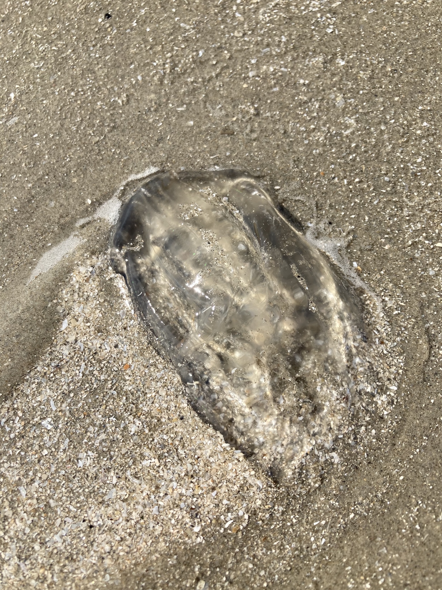
The sea walnut doesn’t look that magical when washed ashore. Photo by iNaturalist user twosandcastles.**
One interesting phenomenon in the sea walnut is its defecation. It has a sack-like gut that most of the time has only the mouth as its opening. However, when its gut is filled with feces, part of it kind of balloons out until touching the epidermis and fuses with it, forming a temporary anus through which feces are expelled. The process is reverted and the anus disappears soon after. But there is one more peculiar thing about this story. Defecation occurs at regular intervals, about once every hour in the adults and once every 10 minutes in the larvae. Can you imagine that? Having to make and unmake your anus every hour? Or every ten minutes?
Despite its bioluminescence and iridescent colors, the sea walnut has a dark side as well. As I said above, this species is native to the Western Atlantic, where it lives just fine with other sea creatures. In the 1980s, however, it was accidentally introduced in the Black Sea, probably through ballast water from merchant ships. First observed in 1982, the species reached a density of up to 400 specimens per m³ in 1989. Its presence caused a dramatic drop in the populations of an anchovy species, Engraulis encrasicholus, a commercially important fish in this region. To control its population, another comb jelly was deliberately introduced in the Black Sea, Beroe ovata, which is a natural predator of the sea walnut. Fortunately, both species seem to have reached a stable predator-prey dynamic, otherwise the situation could have become even worse.
But humans never get tired of finding new ways to ruin ecosystems, right? Russia developed a network of channels running across the country’s rivers that connect several saltwater bodies for navigation, including the Black Sea, the Caspian Sea, the Baltic Sea, and the White Sea. As a result, the sea walnut was able to spread from the Black Sea into the Caspian Sea in 1999. There, it started to feed on the eggs and larvae of small fish and led to a reduction in the population of larger fish and seals.
During the 21st century, the sea walnut continued to spread across European seas, colonizing the Mediterranean, the Baltic and the North Seas. Its impact on these areas is still unknown, but it could be catastrophic, especially in the Baltic Sea, which is one of the most impacted marine environments in Europe.
Schnitzler, C. E., Pang, K., Powers, M. L., Reitzel, A. M., Ryan, J. F., Simmons, D., … & Baxevanis, A. D. (2012). Genomic organization, evolution, and expression of photoprotein and opsin genes in Mnemiopsis leidyi: a new view of ctenophore photocytes. BMC biology, 10(1), 1-26. https://doi.org/10.1186/1741-7007-10-107
Shiganova, T. A., Sommer, U., Javidpour, J., Molinero, J. C., Malej, A., Kazmin, A. S., … & Delpy, F. (2019). Patterns of invasive ctenophore Mnemiopsis leidyi distribution and variability in different recipient environments of the Eurasian seas: A review. Marine environmental research, 152, 104791. https://doi.org/10.1016/j.marenvres.2019.104791
However, in 2018, one marine polyclad seems to have decided to explore the world outside the oceans by riding a toad in Bangladesh. How did this happen? Well, no one is sure. A group of researchers working on Nijhun Dwip Island found a common Asian toad (Duttaphrynus melanosticus) walking through an agricultural field with a polyclad attached to its dorsum.
The toad and the polyclad. A new Aesop’s fable. Credits to Rabbe et al. (2020).*
A canal that passes through the field can be flooded by the sea during high tide and this is how the researchers think the flatworm ended up on land and eventually on the back of the toad. Unfortunately, because the toad was spotted by the researchers, the poor polyclad ended up collected and killed and is now preserved in a lab.
Rabbe MF, Roy DK, Mohammad N, Liza FT, Mukutmoni M, Alam MM, Begum A & Jaman MF. 2020. A Novel Natural History Phenomenon: A Free-living Marine Flatworm (Polycladida) Attached to a Common Asian Toad (Duttaphrynus melanostictus). Reptiles & Amphibians 27: 293–294. https://journals.ku.edu/reptilesandamphibians/article/view/14404


 This work is licensed under a Creative Commons Attribution 4.0 International License.