Friday Fellow: Fly-Killing Fungus

by Piter Kehoma Boll

You most likely know about the existence of the parasitic fungi of the family Cordycipitaceae that turn insects and other arthropods into “zombies”. I even presented one here already about 5 years ago, the Chinese caterpillar fungus. But zombie-making fungi did not evolve only once and today we will meet another one that also zombifies insects, more precisely flies.

Its scientific name is Entomophthora muscae and I decided to give it the common name fly-killing fungus. While the most famous zombifying fungi belong to the phylum Ascomycota, the fly-killing fungus belongs to the phylum Entomophtoromycota, which until recently was part of the phylum Zygomycota.

Dead fly of the species Scathophaga stercoraria consumed by the fly-killing fungus. Photo by Hans Hillewaert.*

The fly-killing fungus is actually a complex of species that infects a variety of flies, including house flies. Their life start as an asexual spore, the conidium. When the conidium contacts the surface of a fly, it germinates, forming a germ tube that penetrates the cuticle of the fly. When the germ tube reaches the haemocoel, the cavity in which the fly’s haemolymph (blood) is found, it releases some cells (protoplasts) into the haemolymph. The protoplasts are carried to the brain and grow in areas that control the fly’s behavior. From there, the fungus start to produce hyphae that grow through the whole body, slowly digesting the internal organs. When the fly is about to die, it crawls to a high point in the vegetation or other available structure, stretches the hind legs and opens the wings and eventually dies. Some hours later, the fungus start to produce conidiophores that grow on the surface of the fly. When they are mature, they release conidia into the environment, restarting the cycle.

Dead hoverfly of the species Melanostoma scalare with the conidiophores of the fly-killing fungus coming out of its abdomen. Photo by Wikimedia user TristramBrelstaff.**

The species complex known as Entomophthora muscae can infect a huge variety of flies, including domestic flies, fruit flies, blow flies, mosquitos and many others. As many of the hosts of the fly-killing fungus as medically or economically important species for humans, there have been attempts to “weaponize” this fungus, turning it into a biological control against several fly pests.

This is not an easy task, though. First, the fungus is very sensible to high temperatures and flies can “cure” themselves from the infection by moving to hot environments that inhibit the growth of the fungus. Another problem is that, in order to raise the fungus in the lab, the fungus needs a constant supply of live adults flies.

As in many cases of using biological agents to control pests, there is always the possibility of things going wrong and end up affecting species that were not the original target. The genetic diversity of the fly-killing fungus is not very well known yet, so that we don’t know exactly which fly species each strain (or species) can actually infect.

I hope we don’t end up turning this ecologically innocent fungus into one more villain on the planet due to our misbehavior.

– – –

Like us on Facebook!

Follow us on Twitter!

– – –

References and further reading:

Brobyn, P. J., & Wilding, N. (1983). Invasive and developmental processes of Entomophthora muscae infecting houseflies (Musca domestica). Transactions of the British Mycological Society80(1), 1-8. https://doi.org/10.1016/S0007-1536(83)80157-0

Kramer, J. P., & Steinkraus, D. C. (1981). Culture of Entomophthora muscae in vivo and its infectivity for six species of muscoid flies. Mycopathologia76(3), 139-143. https://doi.org/10.1007/BF00437194

Krasnoff, S. B., Watson, D. W., Gibson, D. M., & Kwan, E. C. (1995). Behavioral effects of the entomopathogenic fungus, Entomophthora muscae on its host Musca domestica: Postural changes in dying hosts and gated pattern of mortality. Journal of Insect Physiology41(10), 895-903. https://doi.org/10.1016/0022-1910(95)00026-Q

Wikipedia. Enthomophthora muscae. Available at < https://en.wikipedia.org/wiki/Entomophthora_muscae >. Access on 12 August 2021.

– – –

*Creative Commons License This work is licensed under a Creative Commons Attribution-ShareAlike 4.0 International License.

**Creative Commons License This work is licensed under a Creative Commons Attribution-ShareAlike 3.0 Unported License.

Leave a comment

Filed under Friday Fellow, Fungi, Parasites

Friday Fellow: Brazilian Rain Lily

by Piter Kehoma Boll

Today species is once again one that was very special to me during my childhood. Its scientific name is either Habranthus robustus or Zephyranthes robusta. I’m not sure which one is currently valid. Its common name in pink rain lily, Brazilian rain lily and many others. It is, however, not a true lily, but a species of the family Amaryllidaceae in the order Asparagales, thus more closely related to things like onions, asparagus and even orchids.

Like most species of Amaryllidaceae and other lily-like monocts, the Brazilian rain lily has an underground bulb that persists through the year. During summer, especially after rainy days, a long scape (stalk), up tp 30 cm long, develops from the bulb and will originate a single flower at its end.

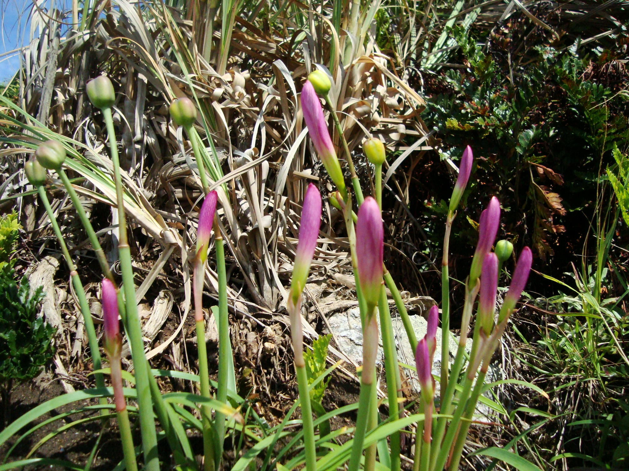
Two beautiful flowers photographed in São Paulo. Photo by Luís Roberto Silva.**

The flowers are up to 6 cm long and have a lavender to pale pink color and a typical lilioid shape, with three petals and three petal-like sepals, as well as six stamens and a single pistil. A single bract (flower-associated leaf) can be seen at the base of the scape. The flowers do not last more than a couple of days and develop into a three-segmented fruit, which will eventually get dry and open to release the seeds.v

Fruits and buds side by side in Penha, Brazil. Photo by iNaturalist user luisfunez**.

Usually after flowering, the plant will produce a group of leaves. These are long, narrow, flat and smooth and are covered by a thin layer of a silverly waxy substance, which gaves them a slightly bluish tinge.

Here you can see the bluish tinge caused by the grayish wax on the leaves. Photo by Krzysztof Ziarnek.*

The Brazilian rain lily is native from southern Brazil and neighboring regions of Argentina, Uruguay and Paraguay but due to its beauty it has become a considerably popular ornamental species and has become naturalized in other parts of the world, especially South Africa and southern United States.

The plant is mildly toxic and can cause irritation in the mucous membranes if ingested. Some alkaloids extracted from the bulbs showed to have acetylcholinesterase- and butyrylcholinesterase-inhibitory activities, which indicates their potential for the development of new drugs for the treatment of dementia.

– – –

Like us on Facebook!

Follow us on Twitter!

– – –

References:

Kulhánková, A., Cahlíková, L., Novák, Z., Macáková, K., Kuneš, J., & Opletal, L. (2013). Alkaloids from Zephyranthes robusta Baker and their acetylcholinesterase‐and butyrylcholinesterase‐inhibitory activity. Chemistry & biodiversity10(6), 1120-1127. https://doi.org/10.1002/cbdv.201200144

Wikipedia. Habranthus robustus. Available at < https://en.wikipedia.org/wiki/Habranthus_robustus >. Access on 4 August 2021.

– – –

*Creative Commons License This work is licensed under a Creative Commons Attribution-ShareAlike 4.0 International License.

**Creative Commons License This work is licensed under a Creative Commons Attribution-NonCommercial 4.0 International License.

Leave a comment

Filed under Botany, Friday Fellow

Friday Fellow: Indo-Pacific Upsidedown Jellyfish

by Piter Kehoma Boll

When we think of a jellyfish, we imagine it pulsating in the water column with its tentacles hanging from its underside. However some jellyfish do not behave exactly like that. This is the case, for example, with those of the genus Cassiopea, which includes the so-called upsidedown jellyfishes. The most studied species of Cassiopea is Cassiopea andromeda, which I decided to call the Indo-Pacific Upsidedown Jellyfish.

As you can imagine from the name I chose to it, the Indo-Pacific upsidedown jellyfish is native from the Indo-Pacific area. The reason why this and other species of this genus are called upsidedown jellyfish is because they are exactly that. They prefer not to swim around like regular a jellyfish, but rather stay near the bottom with their tentacles and mouth facing upward. As a result, they may end up being mistaken for sea anemones.

An adult Indo-Pacific upsidedown jellyfish measures up to 30 cm in diameter and has a brownish bell and arms with tentacles whose shape varies from pointy to flat, round or slender and the color varies from white to brown, red, pink, yellow, green or blue. This species is carnivorous, of course, like most cnidarians, and feeds on small animals that it captures and paralyzes.

The astonishing variety of colors and tentacle shapes in the Indo-Pacific upsidedown Jellyfish. Extracted from Lampert (2016).

Adult Indo-Pacific upsidedown jellyfish are gonochoric, i.e., there are male and female specimens. Males release sperm in the water, which enters the body of the females and fertilizes the eggs. The eggs are kept inside the oral disc (the “mouth”) of the female until they develop into a free-living planula larva. The planula eventually settles when it founds a suitable substrate and develops into a polyp. The polyps can produce buds from their lower portion which detach and end up settling somewhere else to develop into new polyps. As the polyp grows, it turns into a young free-living medusa (ephyra), which grows to become and adult, restarting the cycle. An ideal place for the polyps to settle and develop are the roots of magroves, so this is one of the most common environments to find these jellyfish.

The Indo-Pacific upsidedown jellyfish is also known for its symbiotic relationship with zooxanthellae, unicellular algae (dinoflagellates) of the genus Symbiodinium. It is their presence that gives the jellyfish its brownish color. Both medusae and polyps have the algae inside them, but they are not passed vertically from the mother to the planulae. The polyps, instead, capture them freshly from the environment.

Specimens in an aquarium. Photo by Raimond Spekking.*

The algae provide nutrients for the jellyfish and the jellyfish, in turn, provides protection for the algae and ensures they will receive sunlight for photosynthesis. Several different Symbiodinium species are associated with the polyps, but this number often decreases to a single species in the adult medusae. As the shallow waters in which these jellyfish often live usually are prone to reach considerably high temperatures, not all zooxanthellae can survive.

Although the Indo-Pacific upsidedown jellyfish is native from the Indo-Pacific, it has been introduced in other parts of the world as well. It reached the Mediterranean probably through the Suez canal many decades ago and it has been recently found at the Atlantic coast of the Americas too. Another upsidedown jellyfish is native from this region, the Atlantic upsidedown jellyfish Cassiopea xamachana, and the consequences of both species meeting is still unknown. Let’s hope that the Indo-Pacific invader does not lead the Atlantic species to extinction.

– – –

Like us on Facebook!


Follow us on Twitter!

– – –

References:

Çevik, C., Erkol, I. L., & Toklu, B. (2006). A new record of an alien jellyfish from the Levantine coast of Turkey-Cassiopea andromeda (Forsskål, 1775)[Cnidaria: Scyphozoa: Rhizostomea]. Aquatic Invasions1(3), 196-197.

Hofmann, D. K., Neumann, R., & Henne, K. (1978). Strobilation, budding and initiation of scyphistoma morphogenesis in the rhizostome Cassiopea andromeda (Cnidaria: Scyphozoa). Marine Biology47(2), 161-176. https://doi.org/10.1007/BF00395637

Lampert, K. P. (2016). Cassiopea and its zooxanthellae. In The Cnidaria, past, present and future (pp. 415-423). Springer, Cham. https://doi.org/10.1007/978-3-319-31305-4_26

Morandini, A. C., Stampar, S. N., Maronna, M. M., & Da Silveira, F. L. (2017). All non-indigenous species were introduced recently? The case study of Cassiopea (Cnidaria: Scyphozoa) in Brazilian waters. Journal of the Marine Biological Association of the United Kingdom97(2), 321-328. https://repositorio.unesp.br/bitstream/handle/11449/162535/WOS000395463500012.pdf?sequence=1

Stampar, S. N., Gamero-Mora, E., Maronna, M. M., Fritscher, J. M., Oliveira, B. S., Sampaio, C. L., & Morandini, A. C. (2021). The puzzling occurrence of the upside-down jellyfish Cassiopea (Cnidaria: Scyphozoa) along the Brazilian coast: a result of several invasion events?. Zoologia (Curitiba)37. https://doi.org/10.3897/zoologia.37.e50834

– – –

*Creative Commons License This work is licensed under a Creative Commons Attribution-ShareAlike 4.0 International License.

Leave a comment

Filed under cnidarians, Conservation, Friday Fellow

Friday Fellow: Frog Opalina

by Piter Kehoma Boll

Large multicellular organisms are more than only one individual and actually serve as whole ecosystems to smaller, often single-celled organisms. Among European anurans, including the Europeam common frog presented here last week, a frequent inhabitant of their colon and cloaca is today’s fellow, Opalina ranarum, which I decided to call the frog opalina.

The frog opalina is a unicellular heterokont protist with a flat oval cell that measures up to 400 µm in length and 200 µm in width, so that it is large enough to be seen with the naked eye. It is said that their appearance to the naked eye is of a small opalescent grain, i.e., they have the multicolored aspect of an opal, hence the name of the genus, but I did not find photographs to confirm that. The body is covered by oblique rows of flagella that make it look like a ciliate, although it is not closely related to these protists.

Despite being a single-celled organism, the frog opalina has many nuclei inside its cell, which are often regularly spaced across the cytoplasm. They are all similar nuclei, not forming micro- and macronuclei like in ciliates and other groups.

A live frog opalina. Waves of the beating flagella are visible across the cell, as well as several dots that I think may be the nuclei. Photo by Wikimedia’s user Djpmapleferryman.*

The exact form of interaction that occurs between the frog opalina and its amphibian hosts is unknown. It was for some time regarded as a parasite, but it seems that it is actually more like a commensal, feeding on organic matter presented in the final part of the frogs’ intestine. Therefore, it does not pose a threat to the host, at least not under normal conditions.

Watch some of them moving.

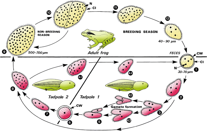
The reproductive cycle of the frog opalina is synchronized with that of their host. Outside of the frogs’ breeding season, the adult frog opalinas live in the posterior end of the frogs’ intestine in the trophozoite or vegetative stage, in which they grow and sometimes reproduce asexually by fission. When spring arrives, they start to show accelerated asexual division, apparently triggered by the frogs’ sex hormones. This accelerated division produce many small individuals with few nuclei that encyst. The cysts are then released in the environment with the frog feces.

Life cycle of the frog opalina. Extracted from Melhorn (2016).

When the frogs’ eggs develop into tadpoles, those end up swallowing some of the released cysts. Inside the tadpoles, the cyst turn back into small trophozoites that continue to divide, becoming smaller and smaller and eventually originate very small individuals that act like gametes. Two of these gametes end up fusing and forming a zygote, which may remain inside the tadpole or be released into the environment to be swallowed again by another tadpole. Regardless of which path the zygote take, it will slowly develop into a new adult trophozoite as the tadpole grows to become an adult frog too, which restarts the cycle.

Although the frog opalina is more often depicted as living inside the European common frog, it can be found in the intestine of other European anurans as well. Are they all really the same species, though? We probably need more genetic studies on these protists’ populations to find the answer.

– – –

Like us on Facebook!

Follow us on Twitter!

– – –

References:

Mehlhorn, H. (2016). Opalinata. In: Encyclopedia of Parasitology (Ed. H. Mehlhorn). Available at < https://link.springer.com/referenceworkentry/10.1007%2F978-3-662-43978-4_2221 >.

Noblet, G. P., & Yabsley, M. J. (2001). The good and the bad: symbiotic organisms from selected hosts. https://www.researchgate.net/publication/228990244_The_Good_and_the_Bad_Symbiotic_Organisms_From_Selected_Hosts

Olsen, O. W. (1974). Animal Parasites: Their Life Cycles and Ecology.

– – –

*Creative Commons License This work is licensed under a Creative Commons Attribution-ShareAlike 4.0 International License.

Leave a comment

Filed under Friday Fellow, protists

Friday Fellow: European Common Frog

by Piter Kehoma Boll

We are about to reach 300 Friday Fellows and only one amphibian has been presented until now. So I guess it is time to present at last an anuran species and, although there are many interesting species all around the world, I decided to talk about the most common, or at least most known, species, the European common frog, Rana temporaria.

Found across Eurasia, this species is, one could say, the type-species of the anuran amphibians. It still has the very same name that Linnaeus has given to it back in 1758, when all other anurans were also put in the genus Rana.

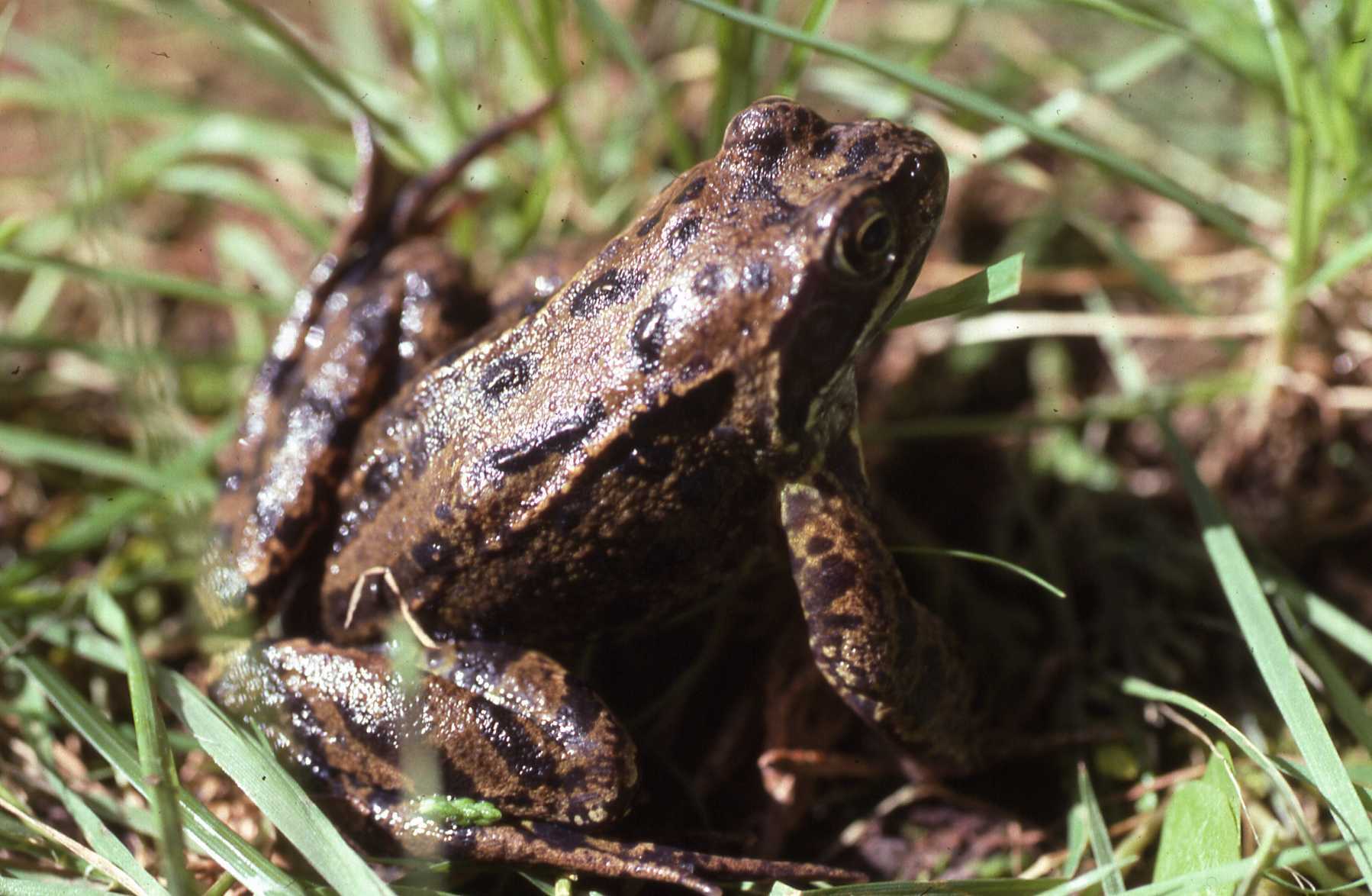
A dark-brown specimen in the UK.

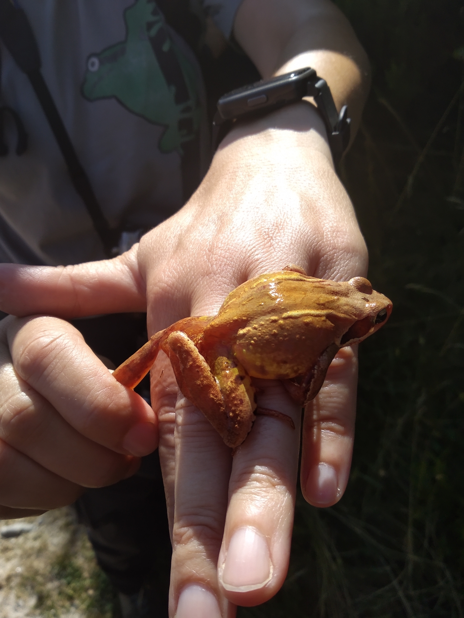
As most true frogs, the European common frog is a semiaquatic species, with adults living their whole life in the water or damp places near waterbodies, especially ponds and marshes. They hibernate during the coldest months of the year, but some populations can remain considerably active even when water is at temperatures very close to freezing. Their skin can vary considerably in color from olive green to several shades of brown and gray, and, in some rare cases, they can even be black or red. They have the ability to adjust their color to their surroundings, thus increasing their camouflage ability. There are also irregular dark blotches distributed across the body, especially on the limbs and flanks.

A yellowish specimen from Spain on a human hand for size comparison.

Adult specimens measure from 6 to 9 cm in length, with females being slightly larger than males. They feed on a variety of invertebrates, including arthropods, gastropods, worms and almost anything small that crawls near them.

In spring, both males and females start to produce sex cells and prepare for mating. They congregate in ponds in large groups and males compete for females through very loud vocalizations and usually those able to produce the loudest and longest calls are preferred. When a female approaches a selected male, he will try to grab her, moving over her, and holding her with his front legs grasping her under her own forelegs. An enarlged, swollen, area on the male’s thumbs, known as the nuptial pad, helps him to hold the female. If other males try to graps her, he kicks them away. The female eventually will release her eggs and he will pour his sperm over them as they are released.

A couple in Switzerland seeking for a nice wet room to make love.

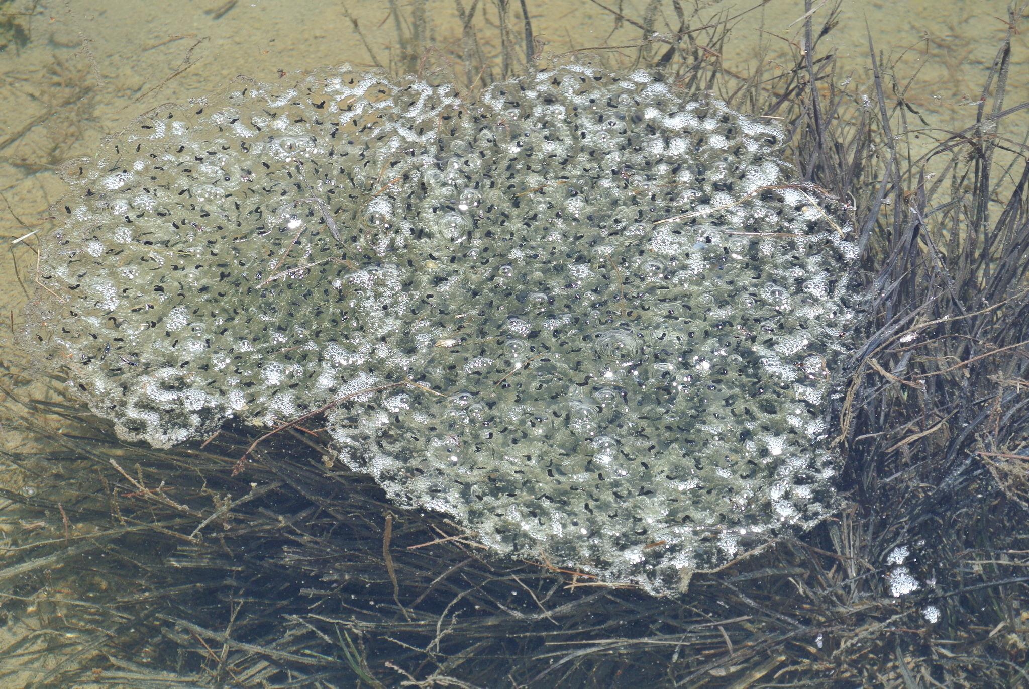
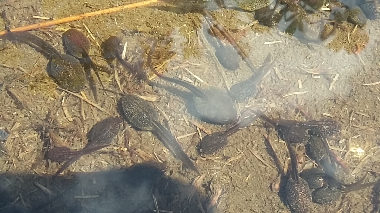
The eggs form jelly-like clusters that float on water and neither the female nor the male will take care of them. The eggs develop and hatch at different rates, depending on temperature. The higher the temperature, the faster the development, and the same happens with tadpoles as they hatch and grow. Newly hatched tadpoles are mostly herbivorous, feeding on algae, but become fully carnivorous with time, eating any small animals they can find, including other, smaller tadpoles.

Lots of eggs floating on a pond in Italy.

The European common frog is usually not exploited by humans as a resource. It is not edible, or at last not often eaten, as far as I can say. It is, however, a very well studied species due to its common occurrence in human-inhabited areas across Eurasia, so that there is a lot of data available about its ecology, genetic diversity, behavior and much more. Its populations are affected by human activities nonetheless, especially urbanization, which causes habitat barriers, and pollution. Nevertheless, it is not a threatened species at the moment.

Tadpoles in Italy.

– – –

Like us on Facebook!

Follow us on Twitter!

– – –

References:

Decout, S., Manel, S., Miaud, C., & Luque, S. (2012). Integrative approach for landscape-based graph connectivity analysis: a case study with the common frog (Rana temporaria) in human-dominated landscapes. Landscape ecology27(2), 267-279. https://doi.org/10.1007/s10980-011-9694-z

Wikipedia. Common frog. Available at < https://en.wikipedia.org/wiki/Common_frog >. Access on 15 July 2021.

Leave a comment

Filed under amphibians, Friday Fellow

Friday Fellow: Iberian Solifuge

by Piter Kehoma Boll

The most popular arachnids are certainly spiders, scorpions and mites, followed by harvestmen. Another interesting but often overlooked group is that of the solifuges, sometimes also known as camel spiders or wind scorpions. This interesting group is characterized by occurring only in deserts and other dry regions of the world.

The species I’m introducing today occurs in dry regions of the Iberian Peninsula. With the scientific name Gluvia dorsalis, this is the only solifuge known to occur in that region, so a common name such as Iberian solifuge seems to be appropriate. Its favorite areas across the peninsula are open grass plains with low vegetation and very low precipitation during summer.

Five pairs of legs? Actually no. Photo by iNaturalist user pepcanto.*

Like all solifuges, the Iberian solifuge looks like a scorpion software forced to run on spider hardware. They look like long spiders with five instead of four pairs of legs. The first pair, however, is not of true legs, but of pedipalps. They are larger than the first three pairs of legs in the Iberian solifuge, but have fewer segments. Solifuges also have huge chelicera compared to other arachnids and can use them as powerful pincers. The legs and pedipalps often have an orange to reddish-brown color, while the abdomen is dark-brown and the prosoma usually reddish, although the color can vary between individuals and across different life stages.

Look at the powerful chelicerae of this female. Photo by Óscar Mendez.*

Female Iberian solifuges are a bit larger than males, reaching up to 2 cm in length, while males reach about 1.7 cm. They are only active during the warm months of the year, between May and November, especially when the environment is very dry. I bet they hate rain almost as much as I hate Bolsonaro.

Males are smaller and have narrower abdomens than females. Photo by Rui Cambraia.*

Being a predator, the Iberian solifuge feeds on a variey of other arthropods, such as all sorts of inects, woodlice and arachnids, including smaller individuals of the same species. They are nocturnal, being active from sunset until about midnight. During the day, males seek shelter under stones, in crevices or among debris, while females and juveniles can dig burrows. Females probably do so to provide a more protected space for thei eggs, while juveniles do so to withstand the winter.

Female feeding on what seems to be a hymenopteran. Photo by iNaturalist user faluke*

Mating occurs between June and August and males die soon after, while females live a bit longer until they lay their eggs, often in a single clutch with more than 100 eggs, although eventually they can lay an additional, but much smaller clutch. The larvae hatch from the eggs about 2 months after they were laid and pass through some instars during the following weeks, becoming juveniles before the temperatures drop and they need to dig their burrows to overwinter. In the next summer they continue their development and overwinter again when they are almost reaching adulthood, which happens at the onset of their third summer.

Female entering her burrow. Photo by iNaturalist user pirataber.**

Because of their spider-like appearance and very rapid movements, solifuges often frighten people, but they are completely harmless tu humans. Different from spiders and scorpions, they do not have any venom. The only harm that they can cause is by biting a human when they are molested but, although painful, the bites are of no medical concern.

– – –

Like us on Facebook!

Follow us on Twitter!

– – –

References:

González-Monliné, A. L., Melic, A., & Barrientos, J. A. (2008) Taxonomía, distribución geográfica e historia natural del endemismo ibérico Gluvia dorsalis (Latreille, 1817) (Solifugae: Daesiidae). Boletín Sociedad Entomológica Aragonesa, 42, 385–395.

Hrušková-Martišová, M., Pekár, S., & Cardoso, P. (2010). Natural history of the Iberian solifuge Gluvia dorsalis (Solifuges: Daesiidae). The Journal of Arachnology38(3), 466-474. https://doi.org/10.1636/Hi09-104.1

Wikipedia. Solifugae. Available at <https://en.wikipedia.org/wiki/Solifugae>. Access on 8 July 2021.

– – –

**Creative Commons License This work is licensed under a Creative Commons Attribution 4.0 International License.

Leave a comment

Filed under Arachnids

Friday Fellow: Common Arcella

by Piter Kehoma Boll

It is time to talk about an amoeba again and one particular group of these protistas thas has not been featured here yet is that of the testate or shelled amoebas. Since, as I already said many times before, it is very hard to find good information of most protists, as they are not as popular as many groups of animals and plants, I will have to present the most “ordinary” of the testate amoebas, Arcella vulgaris, the common arcella.

As most amoebas, the common arcella has an amoeboid body formed by an irregular cell from which pseudopods extend to aid locomotion and prey capture. In the common arcella, though, there is a beautiful and regular shell as well. The shell is formed by chitin, just like the exoskeleton of arthropods or the cell wall of fungi.

A beautiful common arcella seen from above. Credits to Microworld.

When seen from above, its shell has a circular shape and measures about 100 µm (0.1 mm) in diameter. When seen from the side, we can notice that it is actually a dome-shaped structure, having a funnel-shaped concavity on the ventral side. At the deepest part of this concavity there is an opening through which the amoeba can extend its pseudopods and move around. The shell also curves outward at the border of the opening, forming a kind of collar. One could say that the common arcella kind of resembles a microscopic jellyfish.

Lateral view of the common arcella with its jellyfish-like appearance. Photo by Thierry Arnet.*

The common arcella is a freshwater species and can be found in lakes, shallow ponds or even in water-soaked soil. Its diet consist of small single-celled organism, including bacteria, algae and fungi. Bacteria, however, are the most important item in its diet, as laboratory experiments has show that its development is impaired when raised on only algae, only fungi or a combination of algae in fungi. On the other hand, it develops normally on a mixed diet that includes bacteria or when it eats only bacteria.

As most species of the genus Arcella, the common arcella has typically two nuclei in its cell. Its reproduction occurs by binary fission and is quite interesting. When it feels that it needs to divide into two, the common arcella retracts is elongated pseudopods into its shell and creates a new, thick and round pseudopod that forms a bulge coming out of the shell opening. It then starts to build a new shell around this bulge, using it as a way to modelate the new shell into its adequate shape. Once the new shell is ready, the nuclei will divide and two of them will migrate into the part of the amoeba that is in the new shell. After that, the cell divides at the point where the two shells are connected and we end up with two amoebas, one living inside the old shell and one inside the new shell. Sometimes the common arcella can also build this new shell and enter as a whole inside it, without dividing in two, and leaving the old shell empty, a phenomenon known as exuviation.

Reproduction of the common arcella. Extracted from Porfírio-Sousa & Lahr (2020).

In some rare occasions the amoeba can also build the new shell and just discard it empty. Why would it do that? No one knows.

In laboratory experiments, when the shell was mechanically removed from the common arcella, the amoebas were able to survive without their shell for some time and eventually build a new one, although this regenerated shell always had an irregular shape and impacted the typical behavior of the amoeba. The process of regeneration the shell takes much more time than building a new one for a daughter cell. While producing a shell for reproduction occurs in only a few minutes, no more than half an hour, regenerating a shell for itself takes up to three days. However, when this regenerated amoeba with a malformed shell divided, the daughter cells had shells of normal shape.

The shell, therefore, is not just a protection for the common arcella, but shapes its whole body and behavior.

– – –

Like us on Facebook!

Follow us on Twitter!

– – –

References:

Laybourn, G., & Whymant, L. (1980). The effect of diet and temperature on reproductive rate in Arcella vulgaris Ehrenberg (Sarcodina: Testacida). Oecologia45(2), 282-284. https://doi.org/10.1007/BF00346471

Pchelin, I. M. (2010). Testate amoeba Arcella vulgaris (Amoebozoa, Arcellidae) is able to survive without the shell and construct a new one. Protistology6(4), 251-257.

Porfírio-Sousa AL, Lahr DJG (2020) Current knowledge and research perspectives of the shell formation process in the genus Arcella (Arcellinida: Amoebozoa). Protistology 14(1): 3–14. https://doi.org/10.21685/1680-0826-2020-14-1-1

– – –

*Creative Commons License This work is licensed under a Creative Commons Attribution-ShareAlike 3.0 Unported License.

Leave a comment

Filed under Friday Fellow, protists

Friday Fellow: Tropical Leatherleaf

by Piter Kehoma Boll

Gastropods show an astonishing diversity of forms, being second only to insects. One of the most diverse groups within them is that of the pulmonate (i.e., lunged) gastropods, but not all pulmonate gastropods actually have lungs. This is the case with the so-called leatherleaf slugs or simply leatherleaves, one of the most peculiar pulmonate families.

Among the leatherleaves, one of the best known species is Laevicaulis alte, often called the tropical leatherleaf in English. This species is native from tropical Africa and, as all leatherleaves, it lacks lungs and has an anus located at the posterior end of the body, different from most pulmonate gastropods, in which it is twisted forward. Adult tropical leatherleaves measure about 8 cm in length and their dorsal side is completely covered by their mantle, which has a dark, almost black tuberculated skin with a central beige line. The tentacles are small and very rarely extend beyond the limits of the mantle, so they are rarely seen from above. The foot, the ventral line with which slugs move, is very narrow, only about 4 to 5 mm wide in adults.

A specimen crawling at night in Mozambique. Photo by iNaturalist user i_c_riddell.**

As most gastropods, the Tropical leatherleaf is a hermaphrodite, but it starts is sexually mature life as a male and later changes its sex to female. They are well-adapted to dry conditions, having a leathery dorsum and a narrow foot, which reduce water loss. Additionally, it can contract into a smaller, rounder shape, which also reduces water loss.

The diet of the Tropical leatherleaf in its native environment is unknown. Living in the leaf litter, it probably feeds on decaying plant matter and small herbs. However, it has been accidentally introduced in many tropical areas around the world, especially in Australia and surrounding islands in the Pacific, such as Taiwan, Hawaii, Fiji, New Caledonia and Samoa, as well as in India and Indonesia. There, it can become a serious pest for some crops, such as tomatoes, cucumbers and spinach.

A specimen in Taiwan. Photo by Beren Tofino.*

More than a pest to plantations, this slug can also serve as an intermediate host for the rat lungworm, Angiostrongylus cantonensis, which can cause eosinophilic meningitis in humans and other animals. This is, therefore, one more species that used to be a harmless species in its native environment, but was turned into a villain by human actions.

– – –

Like us on Facebook!

Follow us on Twitter!

– – –

References:

Brodie, G. D., & Barker, G. M. (2012). Factsheet Laevicaulis alte (Ferussac, 1822). http://repository.usp.ac.fj/5436/1/Laevicaulis_alte-_Ferussac-1822.pdf

Wikipedia. Laevicaulis alte. Available at < https://en.wikipedia.org/wiki/Laevicaulis_alte >. Access on 24 June 2021.

– – –

**Creative Commons License This work is licensed under a Creative Commons Attribution 4.0 International License.

Leave a comment

Filed under Conservation, Friday Fellow, mollusks

Friday Fellow: Turkish Towel

by Piter Kehoma Boll

The red algae are the sister group of land plants and one of the most diverse groups of algae. It’s time to introduce one more of them today and I think that Chondracanthus exasperatus is a good choice.

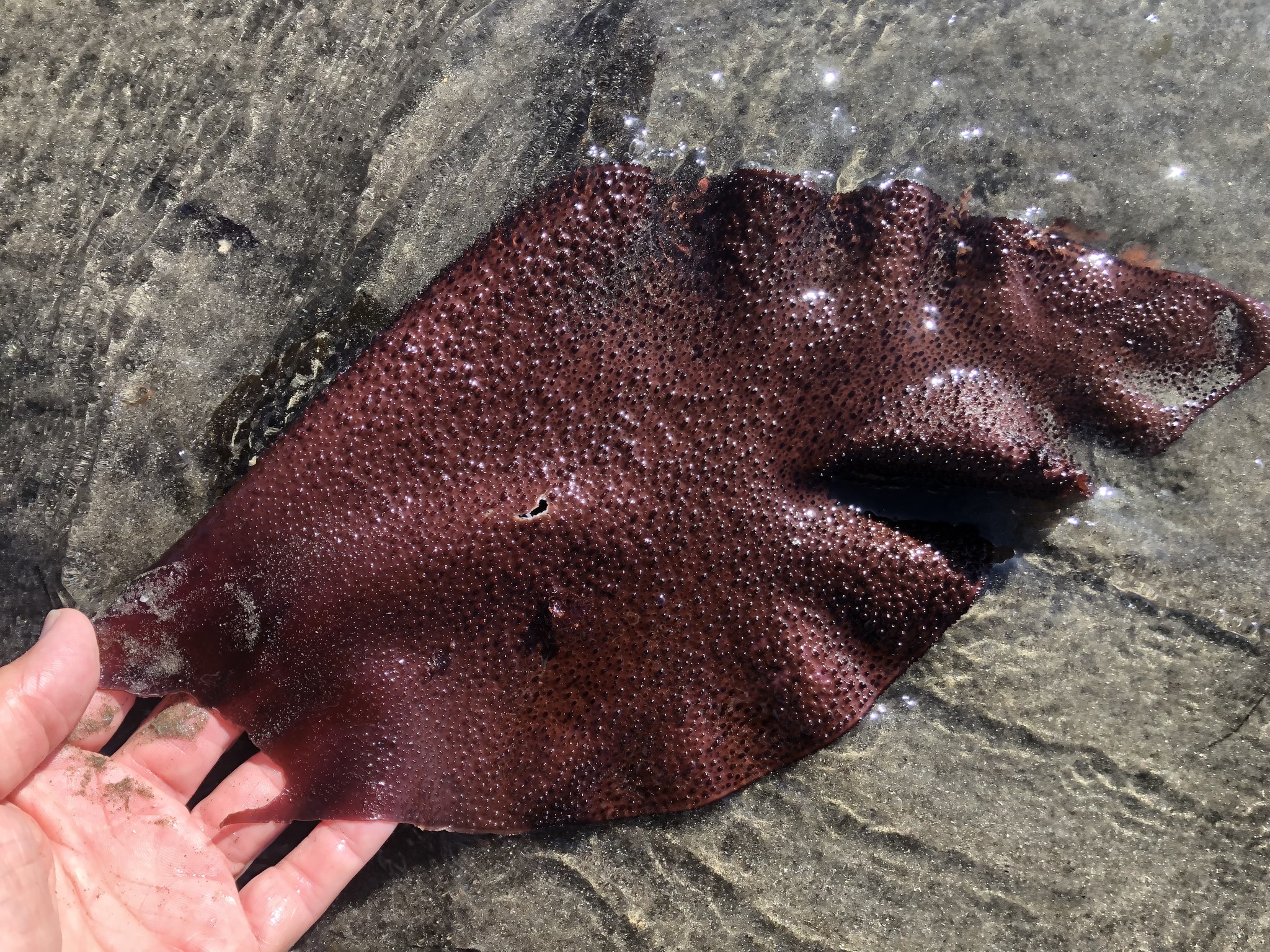
Popularly known as the Turkish Towel, this species is found in the Northern Pacific, especially along the North American coast. It has a leafy thallus, often branched and the surface of the blades have many small papillae, which gives them a rough appearance and make them resemble a towel, more specifically a towel used for hammam or Turkish bath, hence the name. The blades have a greenish color when growing in surface waters, where they get exposed to UV light, but become dark-red to purple when growing in deeper waters. They can grow up to almost 1 m in length and have thick margins marked by rounded teeth.

It really looks like a towel, doesn’t it? Photo by Ryan Durand.*

Little is know about the role of the Turkish towel in the food web. It decomposes very quickly when washed ashore and most detritivorous species seem to avoid eating it. Some herbivorous gastropods, such as the white abalone Haliotis sorenseni, eventually feed on them, but a diet based mostly on the Turkish towel can delay their development or even be fatal.

The Turkish towel is of economic interest to humans because it is a source of carrageenans, which are widely used in the food industry as an alternative to gelatin. Although similar to agar, which is also extracted from red algae and has similar applications, carrageenan is a different compound. The gel found inside the blades have also been used in cosmetic products.

The overall appearance of a blade. Photo by Al Kordesch.

As a result of its usefulness to humans, the Turkish towel is also cultivated and different strategies have been used to find the best way to raise it. Even fast-growing strains have been selected to improve productivity but, overall, the techniques still need improvement.

However, it is necessary to highlight that the safety of consuming carrageenans is not yet well supported by research, so beware! And considering how many marine species avoid eating the Turkish towel, we should proceed with caution with this species as well.

– – –

Like us on Facebook!

Follow us on Twitter!

– – –

References:

Merrill, J. E., & Waaland, J. R. (1979). Photosynthesis and respiration in a fast growing strain of Gigartina exasperata (Harvey and Bailey). Journal of Experimental Marine Biology and Ecology39(3), 281-290. https://doi.org/10.1016/0022-0981(79)90131-X

Sylvester, A. W., & Waaland, J. R. (1983). Cloning the red alga Gigartina exasperata for culture on artificial substrates. Aquaculture31(2-4), 305-318. https://doi.org/10.1016/0044-8486(83)90321-6

Waaland, J. R. (2002). Recirculating Culture for Chondracanthus Exasperatus. Journal of Phycology38, 36-36. https://doi.org/10.1046/j.1529-8817.38.s1.102.x

Wikipedia. Chondracanthus exasperatus. Available at <https://en.wikipedia.org/wiki/Chondracanthus_exasperatus>. Access on 17 June 2021.

– – –

*Creative Commons License This work is licensed under a Creative Commons Attribution 4.0 International License.

Leave a comment

Filed under Algae, Friday Fellow

Friday Fellow: Coldwater Disease Bacterium

by Piter Kehoma Boll

Bacteria are some of the most diverse organisms on Earth and they perform all sort of ecological roles, although they are more often associated with diseases by the average human being. This is, of course, due to the fact that most bacteria that have a direct and perceptible influence on human life are, in fact, pathogenic, often parasitic, bacteria. Today I am introducing one of those pathogenic bacteria, but not one that infects humans.

Currently known as Flavobacterium psychrophilum, this species is known to infect freshwater fish, causing a disease known as bacterial coldwater disease (CWD). As a result, I will call this species the coldwater disease bacterium, or CWD bacterium for short.

The typical aspect of CWD bacteria seen under the microscope. Extracted from Cipriano & Holt (2005).

The CWD bacterium is a typical rod-shaped bacterium measuring up to 1 µm in width and 5 µm in length. It lacks any type of flagellum or other structure that helps it move, but it can move by gliding, although this movement is incredibly slow and very difficult to observe. When cultured in a growth medium, they produce small 3-mm-diameter yellow colonies with thin margins.

Several colonies of CDW bacteria on a culture medium. Credits to Eva Säker (SVA) & Karl-Erik Johansson (BVF, SLU & SVA).*

Living in freshwater, the CWD bacterium prefers cold waters, with temperatures of 16 °C or lower, with the optimal temperature being 13°C. They grow on all sort of tissues on the body of fish, such as the skin, gills, mucous and internal organs such as the brain, kidney, spleen and the sex organs. Its preferred hosts are salmonoid fishes, such as salmons and trouts, but it can be found in other species eventually.

The CWD bacterium is an aerobic bacterium but is unable to use carbohydrates as a source of carbon, feeding on peptides instead. Thus, it secretes enzymes on the host’s tissues to degrade its proteins into peptides, causing structural damage.

Infected fish show tissue erosion, which often begins on the caudal fin and eventually spread. Fins become dark, ragged, split or torn and may be completely lost. Ulcerations appear on the skin, especially around the jaw, and the fish present behavioral issues such as spiral swimming and lethargy. The infection often kills the fish but sometimes a milder chronic infection can occur, which, however, still causes considerable behavioral changes in the host.

Lesions caused by Flavobacterium psychrophilum in the rainbow trout Oncorhynchus mykiss (A) and the coho salmon Oncorhynchus kisutsch. Extracted from Starliper et al. (2011).

The bacteria are often transmitted from fish to fish via direct fish contact, but infected adult fish can end up passing the infection directly to their offspring through infected eggs. The infection can be treated in early stages using the antibiotic oxytetracline or by adding quaternary ammonium cations to the water.

In natural environments the problems caused by this infection are rarely problematic and its damage is more often seen in fish farms, where the poor creatures are forced to live in higher densities, which increases the bacterium’s success. Apparently native to North America, where it was discovered in the 1940s, it was spread via fish farming across the whole world in the following decades. We humans, therefore, are once more the main reason why this species has become a worldwide problem.

– – –

References:

Cipriano, R. C., & Holt, R. A. (2005). Flavobacterium psychrophilum, cause of bacterial cold-water disease and rainbow trout fry syndrome. Kearneysville, WV: US Department of the Interior, US Geological Survey, National Fish Health Research Laboratory.

Langevin, C., Blanco, M., Martin, S. A., Jouneau, L., Bernardet, J. F., Houel, A., … & Boudinot, P. (2012). Transcriptional responses of resistant and susceptible fish clones to the bacterial pathogen Flavobacterium psychrophilum. PLoS One7(6), e39126. https://doi.org/10.1371/journal.pone.0039126

Starliper, C. E. (2011). Bacterial coldwater disease of fishes caused by Flavobacterium psychrophilum. Journal of Advanced Research2(2), 97-108. https://doi.org/10.1016/j.jare.2010.04.001

Wikipedia. Flavobacterium psychrophilum. Available at < https://en.wikipedia.org/wiki/Flavobacterium_psychrophilum >. Access on 10 June 2020.

– – –

*Creative Commons License This work is licensed under a Creative Commons Attribution 2.5 Sweden Generic License.

1 Comment

Filed under Bacteria, Friday Fellow, Parasites