Monthly Archives: July 2022

The richest is not always the commonest: a lesson from flowering plants in a Cerrado outcrop

by Piter Kehoma Boll

The complexity of ecosystems is sustained by a variety of relationships that different species have with each other and that are often adapted to the environment in which they live. Although we usually think of relationships based on conflicts, such as predation, parasitism, and competition, beneficial relationships are almost as important and common, especially when we think of flowering plants, as many plant species rely on animals to pollinate them and disperse their seeds.

The different ways through which plants are pollinated are called pollination syndromes and include anemophily (pollination by wind), melittophily (by bees), phalenophily (by moths), sphingophily (by hawk moths), psychophily (by butterflies), myophily (by flies), cantharophily (by beetles), chiropterophily (by bats) and ornithophily (by birds), and there are generalist plants as well, whose flowers can be pollinated by several different animals. Now considering the way plants have their seeds dispersed, the classification is usually into only three categories: zoochory (by animals), anemochory (by wind), and autochory (by the plant itself, because why wait for animals or the wind? Ain’t nobody got time for that!).

Birds are among the many animals that can pollinate flowers (A) and disperse seeds (B). Image by Cássio Cardoso Pereira.*

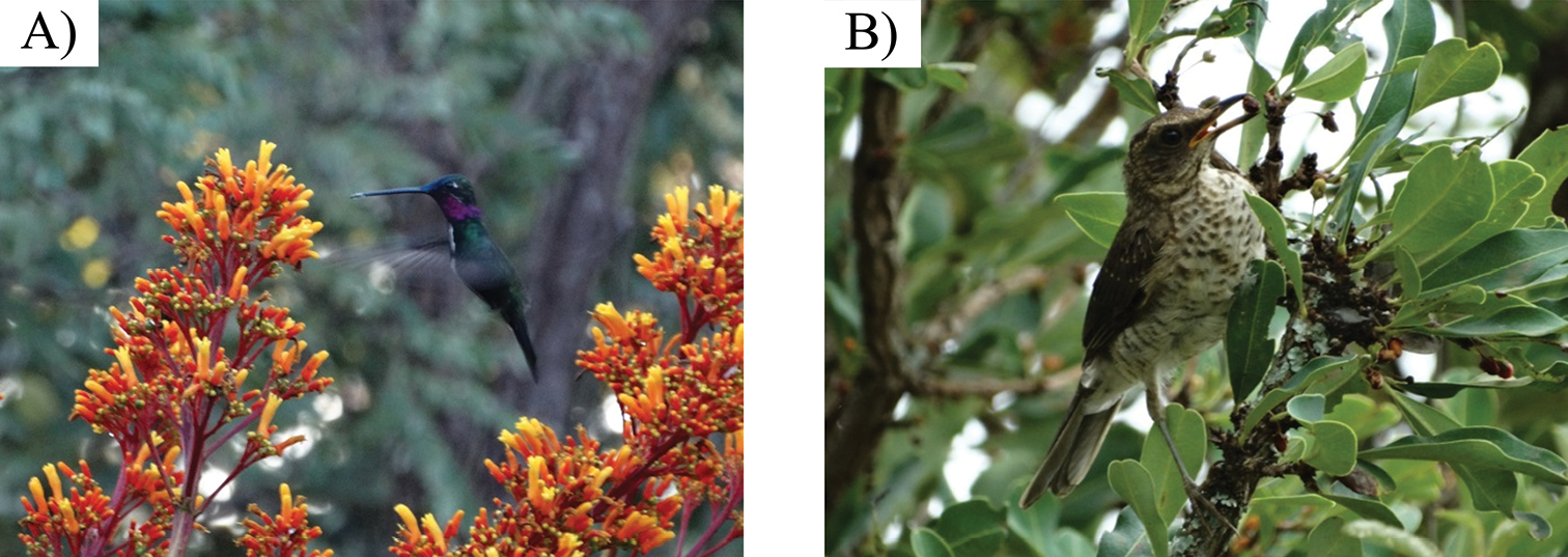
We often think of bees and butterflies as the most common pollinators. Indeed bees are by far the most common and important, but actually very few plants rely exclusively on butterflies for pollination. Flies, beetles, moths, and even birds and bats often pollinate more plant species in a given ecosystem. Regardless of the ecosystem being a dense forest, an open grassland, or a shrubby savanna, bees are always the ones doing the job for most plant species.

Now regarding seed dispersal, the configuration of the ecosystem is much more important and causes drastic changes in the frequency of dispersal syndromes. In open areas such as grasslands and savannas, anemochory is often considered to be the predominant dispersal syndrome. In forests, however, zoochory would dominate, as there is not enough wind to blow seeds around.

When we survey the dispersal syndromes in forests, we find that most plant species have, indeed, their seeds dispersed by animals. However, a survey in grasslands and savannas can show results that look puzzling at first. Sometimes all three dispersal syndromes occur in the same proportion and sometimes lots of species continue to be dispersed by animals, while the wind is important only to a few. Were we wrong in our predictions then? Not necessarily.

One problem is that most studies, almost all actually, only compare pollination and dispersal syndromes by the number of species in that area. However, plant species are not evenly distributed in the environment. Some species have lots of individuals, being dominant in their ecosystems, while others occur in a much smaller number. Does the proportion of dispersal syndromes remain the same if we consider the number of individuals and not species? Not necessarily.

A recent study evaluated the pollination and dispersal syndromes of plants in an area of the Brazilian Cerrado biome, more specifically an area of Cerrado Rupestre (one of the less known Cerrado physiognomies). The researchers not only considered the distribution of the syndromes according to the number of species but also according to the number of individuals. Most plant species were pollinated by bees, as expected, and most individuals were pollinated by bees as well. However, while most species had their seeds dispersed by animals, most individuals had their seeds dispersed by wind. This means that, although most species rely on animals to disperse seeds, they tend to occur in a lower density, with fewer individuals per area. On the other hand, wind-dispersed species have a very high density, so most individuals in an area belong to them.

When we consider pollination and dispersal syndromes according to species or individuals, the picture can change drastically. Although most species are dispersed by animals in this Cerrado fragment (B), most plant individuals actually belong to species dispersed by wind (D). Credits to Pereira et al. (2022).*

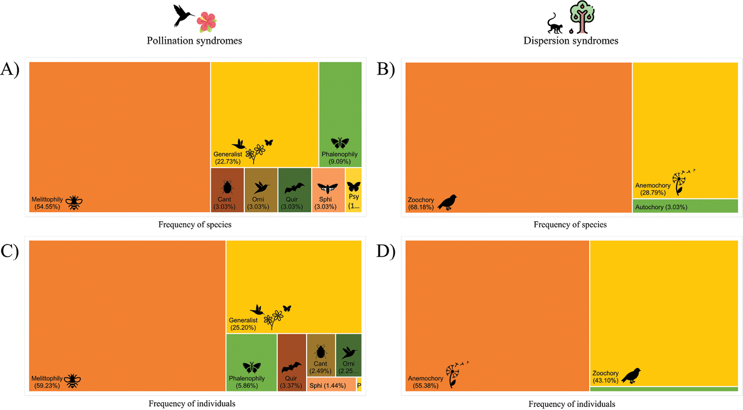
When we consider the distribution of dispersal syndromes only according to species, the results seem to contradict what is expected for a savanna, but looking at it from the perspective of individuals makes it clear that the pattern follows the predictions.

Being aware of this is important for several reasons, especially to allow adequate management programs to protect such areas. The stability of an ecosystem does not depend solely on the species richness but also on the abundance of each species. By analyzing the distribution of dispersal syndromes from both perspectives, we can see that the wind is the main disperser for this ecosystem as a whole, but animals are still important dispersers to keep the species richness high and, in turn, a high richness of plant species is important to sustain the animal species. This makes our understanding of the whole system very different from what we would know from data on species alone. Now let’s hope future studies will start to address this issue from both perspectives as well.

– – –

Follow us on Twitter!

– – –

References:

Kuhlmann, M., & Ribeiro, J. F. (2016). Evolution of seed dispersal in the Cerrado biome: ecological and phylogenetic considerations. Acta Botanica Brasilica30, 271-282. https://doi.org/10.1590/0102-33062015abb0331

Pereira, C. C., Arruda, D. M., Soares, F. D. F. S., & Fonseca, R. S. (2022). The importance of pollination and dispersal syndromes for the conservation of Cerrado Rupestre fragments on ironstone outcrops immersed in an agricultural landscape. Neotropical Biology and Conservation17(1), 87-102. https://doi.org/10.3897/neotropical.17.e79247

– – –

*Creative Commons License This work is licensed under a Creative Commons Attribution 4.0 International License.

Leave a comment

Filed under Botany, Ecology, Entomology

Jaguars help deer eat bones, or: the food web is wild!

by Piter Kehoma Boll

We get used to thinking of the food chain, or more precisely the food web, as something considerably regular and flowing in a single direction: plants synthesize nutrients by photosynthesis, herbivores eat plants, carnivores eat herbivores and stuff. But nature does not work as regularly as we think and sometimes things can look very crazy.

Although many animals are strictly herbivores indeed, this is not exactly the case for many ungulates that we may see as vegan, such as ruminants. It is actually not that rare to find ruminants chewing on bones of dead animals, sometimes when they still have some flesh attached to them. The main reason for herbivores to eat bones is to acquire minerals, such as calcium, which are important not only for growing bones and horns or antlers but also for conducting nerve impulses and other biochemical roles.

Now observations with a single camera trap in Santa Rosa National Park in Costa Rica recorded more than a hundred events of white-tailed deer (Odocoileus virginianus) chewing on bones of turtle carcasses on a beach. The deer included males with growing antlers, lactating females, and growing fawns, all of which need additional amounts of calcium to grow antlers, replace calcium lost in milk production, and grow bones, respectively.

Ok, those deer eat a lot of turtle bones, so what? Well, the reason why there are so many bones available for them to feast on is that jaguars prey on turtles that come to that beach to lay their eggs. Jaguars are among the most important predators in the Neotropics and deer are obviously one of their prey. While the presence of jaguars can be a threat to the life of the white-tailed deer, they are also essential to provide the bones on which the deer feed to get healthy.

Jaguars eat turtles on the beach and let their bones behind. Later, deer come to feed on the bones that their main predator left as a gift. “From jaguar to dear, with love, so that you can grow healthy and feed me later”. Credits to Morera et al. (2022).*

This almost turns the food web upside down, right? Or at least add some very weird loops in it.

– – –

Follow us on Twitter!

– – –

Reference:

Morera, B., Montalvo, V., Sáenz-Bolaños, C., Cruz-Díaz, J. C., Fuller, T. K., & Carrillo, E. (2022). Osteophagia of sea turtle bones by white-tailed deer (Odocoileus virginianus) in Santa Rosa National Park, northwestern Costa Rica. Neotropical Biology and Conservation17(2), 143-149. https://doi.org/10.3897/neotropical.17.e87274

– – –

*Creative Commons License This work is licensed under a Creative Commons Attribution 4.0 International License.

Leave a comment

Filed under Behavior, Ecology

Friday Fellow: Common Tusk Shell

by Piter Kehoma Boll

Mollusks make up the most speciose animal phylum after the arthropods, but only three of its classes are popular among the general public. One of the less known classes is Scaphopoda, which includes a group of mollusks known as tusk shells. Among the about 500 described species, one is named Antalis entalis, known as the common tusk shell.

The common tusk shell is found in the waters of the North Atlantic and, like all tusk shells, it spends most of its life buried in the sediments of the sea floor. Its body measures about 4 cm in length and is surrounded by an elongated shell that has an opening at each end, one end being narrower than the other. The wider opening is the one through which it extends its foot and a series of small tentacles known as captacula. Tusk shells lack eyes completely, at least as adults. Their free-swimming larvae have a brief stage in which very simple eyes occur.

Most of the body of the common tusk is permanently surrounded by the shell. Only its foot and very small tentacles, the captacula, are extended through the larger opening, as seen in the first shell of this image.

Due to the weird anatomy of tusk shells, it is hard to tell which part of their body is the anterior end or posterior end, or which is the dorsum and which is the venter, but this wider opening is often considered the ventral side or anterior end. The narrower opening at the other end of the shell is directed upward and is mainly used for respiration. Water enters through this opening, bringing oxygen, and leaves carrying carbon dioxide away.

As adult common tusk shells never leave the sediment voluntarily, they feed on other organisms that live on the same substrate, with foraminifers making up a large portion of their diet. They use the captacula to capture their prey and ingest them. The feces are eliminated through the anus, which is located at the same wider opening of the shell.

Although about 500 hundred extant species of tusk shells have been described, we know very little of their ecology and it is even hard to find photographs of living specimens. The common tusk shell is one of the most studied species, but these studies are related to its anatomy, distribution, and embryonic development, as well as phylogenetic studies. The importance of tusk shells to marine ecosystems is largely unknown.

– – –

Follow us on Twitter!

– – –

References:

Antalis entalis (Linnaeus, 1758) in GBIF Secretariat (2021). GBIF Backbone Taxonomy. Checklist dataset https://doi.org/10.15468/39omei accessed via GBIF.org on 2022-07-28.

Reynolds, P. D. (2002). The scaphopoda. Advances in marine biology42, 137-236. https://doi.org/10.1016/S0065-2881(02)42014-7

Wollesen, T., McDougall, C., & Arendt, D. (2019). Remnants of ancestral larval eyes in an eyeless mollusk? Molecular characterization of photoreceptors in the scaphopod Antalis entalis. EvoDevo10(1), 1-12. https://doi.org/10.1186/s13227-019-0140-7

Leave a comment

Filed under Friday Fellow, mollusks, Zoology

Friday Fellow: Red Foram

by Piter Kehoma Boll

In the warm waters of the North Atlantic, especially in the Caribbean Sea and the Gulf of Mexico, we can find one of the most amazing foraminifers. Its scientific name is Homotrema rubrum and it is sometimes called the red foram.

This is once again a large foraminifer, easily visible to the naked eye, although not as large as some other species. They are a sessile, encrusting foraminifer that grows on the surface of rocks, shells and corals, and you can detect them as small, often irregular red plates measuring a few millimeters in size on the surface of hard substances. In some places, such as Bermuda, they are so abundant that the sand of the beaches acquired a pink color due to the huge number of skeletons of dead red forams washed ashore.

Some red forams growing on a coral reef in Bermuda, one of the most iconic places where this species is found in abundance. Photo by Ben Eddy.*

Like all foraminifers, the red foram is a single-celled organism and, although some sources that I consulted stated that it is a colonial species, I understood that every red “plate” is actually a single cell, although sometimes you can find several of them nearby and even a bit overlapped.

A piece of dead coral covered by numerous skeletons of dead red forams in Bonaire, with a human hand for size comparison. Photo by Ali and Brice.*

The red shell that surrounds the cell of the red foram can have one or many openings through which this lovely protist extends its reticulopodia—the long, narrow, branching and anastomosing pseudopodia—to capture prey. For some time it was thought that the main, and perhaps only, food of the red foram were small unicellular algae, but now it is known that they feed on other organisms as well, including some small crustaceans. They are true predators!

But there are other surprises about this little fellow. To help it capture its prey, it collects sponge spicules (the small needle-like structures that form the skeleton of sponges) and embeds them into its own cell, using them to improve the support of its reticulopodia. It is almost like a single-celled organism using tools to eat! (Ok, this may be a little bit of a stretch).

In this specimen from Florida, USA, you can see the collected sponge spicules appearing as several small sets of needle-like structures. Photo by Jeff Goddard.*

Like other encrusting foraminifers, the red foram likes to grow inside cracks, crevices and other spaces of coral reefs. As its shell consists of magnesium calcite, it helps reinforce the coral framework and provides carbonate for the corals to grow as well. Thus, they are very important to make the reefs more resistant to climate change since the raise in CO2 levels is making the oceans more acidic, which increases coral reef erosion.

– – –

Follow us on Twitter!

– – –

References:

Elliott, J. M., Logan, A., & Thomas, M. L. H. (1996). Morphotypes of the foraminiferan Homotrema rubrum (Lamarck): distribution and relative abundance on reefs in Bermuda. Bulletin of Marine Science58(1), 261-276. https://www.ingentaconnect.com/content/umrsmas/bullmar/1996/00000058/00000001/art00016

Phalen, W. G. (2015). Homotrema rubrum (Lamarck): distribution and biology of a potential reef bioindicator and underwater angler (Doctoral dissertation, University of Georgia). https://esploro.libs.uga.edu/esploro/outputs/graduate/Homotrema-rubrum-Lamarck-distribution-and-biology/9949333258902959

– – –

*Creative Commons License This work is licensed under a Creative Commons Attribution-NonCommercial 4.0 International License.

Leave a comment

Filed under Friday Fellow, protists

The secret sex life of the bloody ribbon worm

by Piter Kehoma Boll

Ribbon worms (phylum Nemertea) are an interesting but understudied animal phylum. It includes many marine species, such as the bootlace worm, Lineus longissimus, the longest known animal. One of its close relatives is the bloody ribbon worm, Lineus sanguineus, which is much shorter but found in oceans worldwide.

A bloody ribbon worm in New Zealand. Photo by Amaya M.*

A peculiar aspect of the bloody ribbon worm is the fact that no one has ever seen them having sex, I mean, reproducing sexually. Marine ribbon worms just throw their gametes in the water and do not have sexual intercourse per se. Anyway, while most ribbon worms, including other species of the genus Lineus, have sexual reproduction as their main reproductive strategy, the same is not true for Lineus sanguineus, or is it? After decades of observations, no one has ever seen this species reproducing sexually, even though males and females have been seen producing sperm and eggs. However, females produce very few eggs compared to other species and seem to “abort” them. The main mode of reproduction of this species is by fragmentation and later regeneration of the missing parts, similar to how many planarians do. This is, in fact, very rare among ribbon worms.

But do they really live without sexual reproduction? A recent study by Christina Sagorny and Jörn von Döhren challenged this idea. They compared the sequences of three genes from individuals from different populations around the world. If asexual reproduction were the only form of reproduction, the expected pattern would be of little genetic variation within populations and more genetic variation between populations. The results of the haplotype networks, however, show otherwise. There is a huge genetic variation within populations and little variation between populations, which suggests not only that sexual reproduction occurs but that the species constantly spreads around the world, possibly via the planktonic larvae.

Haplotype networks of the bloody ribbon worm. If asexual reproduction was the only form of reproduction, one would expect fewer haplotypes per region and few shared haplotypes between regions, so that the circles would have only one or a few colors. Extracted from Sagorny & von Döhren (2022).

By raising some worms in the lab under summer conditions (18° C and 16 h of light) and others under winter conditions (9° C and 8 h of light), they found that most worms become sexually mature during winter and, surprisingly, reproduce by fission more often in winter too. More than that, they even found some larvae in the water, indicating that sexual reproduction occurred successfully.

What can we conclude? The bloody ribbon worm does have sex. It just likes to make it in private.

– – –

Follow us on Twitter!

– – –

Reference:

Sagorny, C., & von Döhren, J. (2022). Occasional sexual reproduction significantly affects the population structure of the widespread, predominantly asexually reproducing marine worm Lineus sanguineus (Nemertea: Pilidiophora). Marine Biology169(7), 1-17. https://doi.org/10.1007/s00227-022-04077-0

– – –

*Creative Commons License This work is licensed under a Creative Commons Attribution-NonCommercial 4.0 International License.

Leave a comment

Filed under genetics, Molecular Biology, worms, Zoology

Friday Fellow: Brazilian Diamond Weevil

by Piter Kehoma Boll

Weevils are the largest family of beetles, which are the largest order of insects, which are the largest class of arthropods and so on… Thus, it is time to bring one more weevil here. I decided to talk about a beautiful and considerably popular Brazilian species, Entimus imperialis, which is known as the Brazilian Diamond Weevil.

Occurring in the Atlantic Forest in southeastern Brazil, this is a relatively large weevil, measuring up to 3 cm in length. The body has a black background, but it is covered by several rows of pits that are filled with iridescent scales that create an effect of golden green dots due to the presence of photonic crystals. Additionally, there are some iridescent green-blue areas on the legs and forming stripes on the dorsum.

A magnificent specimen from Atibaia, São Paulo. Foto by Whaldener Endo.**

As the scales are arranged inside concave pits, the weevil’s appearance changes drastically depending on the distance from the observer. When seen from far away, it merges perfectly with the green background of the leaves, which is useful to make it invisible to predators. However, when seen from a short distance, the dotted iridescent pattern becomes quite conspicuous and this is thought to be important for the Brazilian Diamond Weevil to recognize others of the same species.

A specimen in Angra dos Reis, Rio de Janeiro. Photo by Edvandro Abreu Ribeiro.*

Despite the extensive study of the optical and structural properties of the scales of the Brazilian Diamond Weevil, little, or perhaps nothing, seems to be known about its ecology. Larvae of other closely related weevil species feed on plants roots while adults feed on the leaves of the same plant, but I was unable to find any information about which plants are used by the Brazilian Diamond Weevil as food.

A specimen from Piranga, Minas Gerais, on a human hand for size comparison. Photo by William R S Maciel.*

According to the records in iNaturalist, adults seem to be found between November and July, especially from November to May. Thus, they most likely die by the end of the autumn and pass the winter as eggs or perhaps larvae.

And that’s all I can tell about this beautiful but little know creature.

– – –

Follow us on Twitter!

– – –

References:

iNaturalist. Brazilian Diamond Weevil (Entimus imperialism). Available at: < https://www.inaturalist.org/taxa/631444-Entimus-imperialis >. Access on 14 July 2022.

Morrone, J. J. (2002). The Neotropical weevil genus Entimus (Coleoptera: Curculionidae: Entiminae): Cladistics, biogeography, and modes of speciation. The Coleopterists Bulletin56(4), 501-513. https://doi.org/10.1649/0010-065X(2002)056[0501:TNWGEC]2.0.CO;2

Mouchet, S., Colomer, J. F., Vandenbem, C., Deparis, O., & Vigneron, J. P. (2013). Method for modeling additive color effect in photonic polycrystals with form anisotropic elements: the case of Entimus imperialis weevil. Optics Express21(11), 13228-13240. https://doi.org/10.1364/OE.21.013228

Wilts, B. D., Michielsen, K., Kuipers, J., De Raedt, H., & Stavenga, D. G. (2012). Brilliant camouflage: photonic crystals in the diamond weevil, Entimus imperialisProceedings of the Royal Society B: Biological Sciences279(1738), 2524-2530. https://doi.org/10.1098/rspb.2011.2651

– – –

*Creative Commons License This work is licensed under a Creative Commons Attribution-NonCommercial 4.0 International License.

**Creative Commons License This work is licensed under a Creative Commons Attribution 4.0 International License.

Leave a comment

Filed under Entomology, Friday Fellow

Friday Fellow: Fungus Root

by Piter Kehoma Boll

Plants are the standard organism when one thinks of photosynthesis, but several species have actually lost the ability to synthesize their own food using light and have become completely heterotrophic. As a result, such plants survive by parasitizing other plants and feeding on their sap. Probably the most famous species of heterotrophic plant is Rafflesia arnoldii, the corpse flower, which has the largest flowers of any plant and was one one our first Friday Fellows almost 10 years ago. But today I will introduce you another, completely unrelated heterotrophic plant, Balanophora fungosa, commonly known as the fungus root.

This species occurs across southeast Asia and Australia, where it lives on the soil and parasitizes the roots of several different plants. As with most heterotrophic plants, the fungus root spends most of its life underground as nothing but a system of roots and rhizomes attached to the host plant. It is only visible on the surface when it produces its flowers, which, like the giant flower of the corpse flower, are also very unusual.

A group of inflorescences coming out of the ground in New Caledonia. We can see the pale bracts and the velvet-like club of female flowers surrounded by the larger male flowers at the base. Photo by iNaturalist user juju98.*

The flowers occur in inflorescences that are actually kind of cute. The overall color varies from pale cream, almost white, to pink. The base of the inflorescence has several bracts (modified, flower-associated leaves) that have the same pale cream to pink color, without any sign of green. The upper part has a club-shaped structure with a velvet-like surface formed by hundreds of tiny female flowers. Surrounding the base of the club are a few male flowers, which are much larger than the female flower, but still very small. The inflorescence as a whole looks similar to some mushrooms, such as puffballs, which is probably the reason for its common name fungus root.

A closeup of an inflorescence in Australia where we can see the male and female flowers in more details. Photo by Aaron Bean.*

I couldn’t find many details about the life cycle of this cute parasite, but it seems to be pollinated by an enormous range of animals, including several types of insects, arachnids and even small vertebrates, which may be attracted to feed on the pollen and nectar or perhaps tricked by the unusual smell that the flowers produce. The smell is unlike the sweet fragrance of most flowers but is also not an unpleasant smell of carrion like that of the corpse flower, the titan arum and so many other plants. Actually, it is said that the flowers smell like a mouse. Perhaps it tricks small mammals to think it is a reproductive member of their species just like some orchids do by mimicking the shape and smell of female bees? This is a possibility, but actually most of the small mammals and birds that visit the flowers are actually nectarivores and are probably only looking for the delicious nectar.

Anyway, there is a lot we still don’t know about this unusual but adorable fungus-like plant.

– – –

Follow us on Twitter!

– – –

References:

Hsiao, S. C., Huang, W. T., & Lin, M. S. (2010). Genetic diversity of Balanophora fungosa and its conservation in Taiwan. Botanical studies51(2). https://ejournal.sinica.edu.tw/bbas/content/2010/2/Bot512-10.pdf

Pierce, R., & Ogle, C. (2017). Musky Rat Kangaroos and other vertebrates feeding from the flowers of the root parasite’Balanophora fungosa’. North Queensland Naturalist47, 14-20. https://search.informit.org/doi/pdf/10.3316/informit.461078578600745

Wikipedia. Balanophora fungosa. Available at < https://en.wikipedia.org/wiki/Balanophora_fungosa >. Access on 8 July 2022.

– – –

*Creative Commons License This work is licensed under a Creative Commons Attribution-NonCommercial 4.0 International License.

Leave a comment

Filed under Botany, Parasites

Friday Fellow: Soybean cyst nematode

by Piter Kehoma Boll

In English, nematodes are known as roundworms, not because they are round like a ball, but because their body is cylindrical like a tube. However, some roundworms are indeed round, or sort of… These include the cyst nematodes of the genus Heterodera, one of which is Heterodera glycines, known as the soybean cyst nematode.

As you may guess by this species having “soybean” in its name, it is a parasite of soybeans. It is, indeed, one of the most devastating pests to attack soybean crops. These worms spend most of their life inside soybean roots, feeding on their tissues.

The typical aspect of the cysts on the roots of an infected plant.

Adults usually measure less than 1 mm in length but males and females are quite different in shape and behavior. Males are longer than females but are very slender, having the typical nematode shape. Once reaching adulthood, they leave the soybean roots and look for females to mate with. Females, on the other hand, have a lemon-shaped, almost spherical body. They never leave the soybean roots but, as they become sexually mature, their bodies swell with eggs, and eventually their posterior end bursts out of the roots, appearing as small white cysts visible to the naked eye.

An egg-filled female. Credits to Agroscope FAL Reckenholz , Swiss Federal Research Station for Agroecology and Agriculture.*

Filled with 200 to 400 eggs, the female dies and turns into a dark cyst. The eggs remain inside her dead body until the environmental conditions are adequate. During this period, the embryo develops from a Juvenile 1 (J1) into a Juvenile 2 stage (J2). The J2 leaves the egg and looks for a new soybean root to penetrate, where it then continues to develop until reaching adulthood.

A scanning electron micrograph showing an egg and a juvenile of the soybean cyst nematode.

The first symptoms of infection by the plant are very subtle, like a mild yellowing of the leaves, which is often mistaken for some nutrient deficiency. The problem is often only noticed during harvest, when yield losses can reach up to 30%.

There are still no effective ways to eliminate the soybean cyst nematode from infected areas. Some parasitic fungi are known to infect the nematode and control its population, and crop rotation is another strategy to reduce the infection load, as planting a different crop for two consecutive years shows a significant reduction in the number of viable eggs in the soil.

Life cycle of the soybean cyst nematode. Credits to George N. Agrios. Extracted from https://www.sciencedirect.com/topics/agricultural-and-biological-sciences/soybean-cyst-nematode

While there are some soybean varieties that are resistant to the infection, there are also different races of the soybean cyst nematode that show different degrees of virulence and new, often more virulent, races are constantly appearing to bypass the resistance developed by the plants. It is evolution being driven right in our farms.

– – –

Follow us on Twitter!

– – –

References:

Texas Invasive Species Institute. Soybean Cyst Nematode. Available at < http://www.tsusinvasives.org/home/database/heterodera-glycines >. Access on 30 June 2022.

Wikipedia. Soybean cyst nematode. Available at < https://en.wikipedia.org/wiki/Soybean_cyst_nematode >. Access on 30 June 2022.

Yan, G., & Baidoo, R. (2018). Current research status of Heterodera glycines resistance and its implication on soybean breeding. Engineering4(4), 534-541. https://doi.org/10.1016/j.eng.2018.07.009

– – –

*Creative Commons License This work is licensed under a Creative Commons Attribution-NonCommercial 4.0 International License.

Leave a comment

Filed under Parasites, worms