Many very small creatures live everywhere, yet we don’t even know that they are there, and even the few of us who do know still know very little about them. One of those creatures is Entomobrya nivalis, commonly known as the cosmopolitan springtail. The fact that it has a common name, however, does not make it a very well studied species, unfortunately.
A specimen of the cosmopolitan springtail in Norway. Credits to Biopix.*
As the common name implies, the cosmopolitan springtail is found all round the world, although it is much more common in the Northern Hemisphere, especially in Europe. Measuring about 2 mm in length, it has an average size for a springtail.
The natural habitat of this little creature are forests, and it may be found both in the leaf litter and on trees. Usually, the first instars, the juveniles, live in the leaf litter, where adults lay their eggs. During summer, adults migrate upward on the trees and live among lichens growing on the branches, a habitat that they seem to consider very cozy.
When winter approaches, and with it the freezing temperatures, the cosmopolitan springtail seeks shelter under lose portions of the bark. This shelter, however, is not enough to protect it from temperatures that would make it freeze to death. As a result, its hemolymph (“blood”) is full of antifreeze compounds that allow it to withstand temperatures as low as -18°C before freezing.
Meier, P.; Zettel, J. (1997) Cold hardiness in Entomobrya nivalis (Collembola, Entomobryidae): annual cycle of polyols and antifreeze proteins, and antifreeze triggering by temperature and photoperiod. Journal of Comparative Physiology B, 167(4): 297–304. https://doi.org/10.1007/s003600050077
Today we celebrate the birthday of the US biochemist and Nobel laureate Julius Axelrod.
Born on 30 May 1912 in New York City, Julius Axelrod was the son of poor Jewish immigrants from Poland, Molly (née Leichtling) and Isadore Axelrod, a basket maker.
After graduating from Seward Park High School, Axelrod studied at the New York University, but after one year his family was not able to afford his studies anymore and he was transferred to the tuition-free College of the City of New York. He graduated in 1933 with a bachelor in Biology. In the same year, he had a laboratory accident when an ammonia bottle exploded, making him lose the sight of his left eye.
Deferred from the World War II draft, Axelrod pursued his dream to become a doctor, applying to numerous medical schools, but was rejected by all of them. He was then hired as a laboratory technician at the Harriman Research Institute at the New York University, preparing buffer solutions and assisting with research on enzymes in malignant tumors. In 1935, the laboratory ran out of fundings and Axelrod lost his position, starting a new work in the Laboratory of Industrial Hygiene at the New York City Department of Health. There, he was assigned the task of monitoring the vitamin content of foods, developing sensitive and specific methods for measuring drugs.
While working there, Axelrod met and married Sally Taub, who had a degree in chemistry from Hunter College, and would eventually have two sons with her.
One day, Axelrod’s department was assigned to clarify why phenacetin and acetanilide, two main ingredients in headache remedies, increased methaemoglobin, a product of haemoglobin’s metabolism, to dangerously high levels in the patients’ blood. In 1946, Axelrod sought assistance of Bernard Brodie, working at the Goldwater Memorial Hospital, to solve this problem. Together they found out that the products of the metabolism of acetanilid and phenacetin, aniline and p-phenetidin, respectively, are the responsible for the elevation of the methaemoglobin’s levels. They also discovered another metabolite of the process, called N-acetyl-p-aminophenol, which is the responsible for the analgesic effects. Today this metabolite is more commonly known as paracetamol, being the world’s most widely used analgesic.
In 1949, Axelrod began to work at the National Heart Institute of the National Institutes of Health. There, he developed research on the metabolism of caffeine, ephedrine and amphetamine in animals, developing an interest on the sympathetic nervous system and its neurotransmitters epinephrine and norepinephrine. He also conducted some of the first experiments on LSD.
In order to advance his career, especially due to the pressure of friends, Axelrod realized that he needed a PhD degree. Thus, he took a year’s leave of absence in 1954, aged 42, to attend George Washington University Medical School. Allowed to submit some of his previous research toward his degree, he was able to receive his PhD one year later, in 1955.
Julius Axelrod in 1973.
In his continuing studies on neurotransmitters, Axelrod discovered that epinephrine and norepinephrine, after released into the synapse, are not inactivated by enzymes, but are rather recaptured by the nerve ending that released them, therefore being recycled. This discovery of the release and reuptake of neurotransmitters made him won the Nobel Prize in Physiology or Medicine in 1970 along with Bernard Katz (1911–2003) and Ulf von Euler (1905–1983). This research later led to the development of selective serotonin reuptake inhibitors, such as fluoxetine (Prozac), a common antidepressant.
Later, Axelrod focused his research on the study of the pineal gland. He and his colleagues showed that the hormone melatonin is generated from tryptophan, which is also the precursor of the neurotransmitter serotonin. They also discovered that the synthesis of melatonin is controled by the circadian rhythm, which is driven by the hypothalamus, and that the hormone has many effects throughout the nervous system, so that the pineal gland works as a biological clock.
Axelrod retired in 1984, but continued to work until he died of a heart attack on 29 December 2004, aged 92.
Homosexual behavior, as you may know, is a widespread phenomenon across the animal kingdom, especially male-male sexual behavior. Like many other aspects of life, this behavior likely evolved independently many, many times and plays different roles in different species.
A study recently published in the journal Animal Behaviour investigated the male homosexual behavior of the red flour beetle, Tribolium castaneum. The team wanted to verify whether a large supply of females or a large number of rival males would influence the occurrence of males having sex with other males.
The red flour beetle, Tribolium castaneum. Credits do CSIRO.*
In order to test that, the researchers directed the evolution of some populations of the red flour beetle in the lab. Some populations were maintained in a male:female ratio of 1:9 and others of 9:1, i.e., in the first group the population consisted of 10% males and 90% females, so males had a greater chance of finding a female than a male and competition between males was very week. In the second group, 90% of the population was made up of males, so females were harder to find and males had to fight for them.
After about 100 generations in which the sex ratios were maintained, the researchers compared the occurrence of male-male sex in both treatments. The results show that males that evolved in an environment where females were abundant and competition between males was low were more likely to engage in homosexual behavior than males that evolved in an environment were competition between males was high and the chances of finding a female were much lower.
The most likely explanation for this difference is that males in highly competitive enviroments need to be better in identifying female individuals to mate with, while in environment where there are plenty of females available, most of the encounters are with females, so the strategy to “have sex with whomever you find” is good enough. The few instances in which such males find other males and mate with them is not enough to reduce their reproductive fitness. In other words, when you live among few females, it is crucial for you to recognize someone as a female and mate with her, otherwise you may end up not passing your genes to the next generation. Now if there are plenty of females you may have sex whenever you want and you certainly will have some children, even if you sometims have fun with your male pals.
Sales K, Trent T, Gardner J, Lumley AJ, Vasudeva R, Michalczyk Ł, Martin OY, & Gage MJG 2018. Experimental evolution with an insect model reveals that male homosexual behaviour occurs due to inaccurate mate choice. Animal Behaviour139: 51–59. https://doi.org/10.1016/j.anbehav.2018.03.004
It’s time for the next diatom to be featured here. Differently from the previous ones, today’s diatom is a freshwater species commonly found in lakes of North America and Eurasia. It has also been reported for South America and Africa, but it is likely that these individuals actually belong to another, closely related species.
Asterionella formosa from a lake of the Rocky Mountain National Park, United States
Named Asterionella formosa, this diatom has small rod-shaped cells that are 60 to 85 µm long and only 2 to 4 µm wide. The individuals usually organize themselves in colonies linked by one of the ends in a star fashion. Most colonies include eight organisms and look somewhat like an asterisk, hence I chose to give the common name asterisk-diatom to the genus, this species then being called the “handsome asterisk-diatom”, from the translation of the specific epithet formosa. However, some colonies may have up to 20 individuals and organize in a more spiral fashion.
A spiral-shaped colony from a small lake in Spain. Credits to Proyecto Agua.*
Originally found and described from water supplies used in London, the handsome asterisk-diatom has a preference for cold waters, occurring commonly in temperate lakes under temperatures between 0 and 15 °C. During summer, when temperatures get too high and the light intensity also increases, its photosynthesis is inhibited by these two factors as well as by the increase in oxygen caused by the metabolism of the species itself as well of other algae from the phytoplanktonic community.
Sexual reproduction is not well known in the handsome asterisk-diatom, but must certainly occur, as asexual reproduction alone leads to a continuous decrease in cell size in all diatoms. Studies on genetic diversity show that this species is very genetically diverse, which proves that sexual reproduction indeed occurs and in a apparently high rate, contributing for the dominance of this species in many of the ecosystems of which it takes part.
De Bruin, A.; Ibelings, B. W.; Rijkeboer, M.; Brehm, M.; Van Donk, E. (2004) Genetic variation in Asterionella formosa (Bacillariophyceae): is it linked to frequent epidemics of host-specific parasitic fungi? Journal of Phycology, 40(5): 823–830. https://doi.org/10.1111/j.1529-8817.2004.04006.x
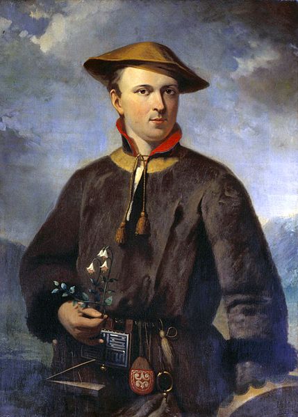
Today we celebrate the birthday of one of the most important figures in the history of biology and certainly the most important one regarding taxonomy, Carl Linnaeus, also known as Carl von Linné or Carolus Linnaeus. There is so much to talk about him, but I will give a very brief summary.
Linnaeus was born in Sweden on 23 May 1707 in a village called Råshult. His father, Nils Ingemarsson, later Nils Linnaeus, was an amateur botanist and a Lutheran minister. Showing an interest in plants since childhood, Linnaeus, during most of his early education, avoided studying other subjects, escaping classes to examine plants in gardens and fields.
In 1724, he entered the Växjö Katedralskola to attend a curriculum designed to those seeking for priesthood, but his progress was not satisfactory. One of his teachers, Johan Rothman, which was also a doctor, suggested that Linnaeus could have a future in medicine and took him to live with his family in Växjö, teaching him physiology and botany. Rothman taught him the basic concepts of botany at the time, including Tournefort’s system to classify plants.
A young Linnaeus in a Laponian costume. Replica of a work by Hendrik Hollander.
In 1728, Linnaeus started to attend Uppsala University by recomendation of Rothman. There he met Olof Celsius, a professor of theology and amateur botanist, who received Linnaeus at his home, which had one of the richest botanical libraries in Sweden. In 1729, Linnaeus wrote a thesis on plant sexual reproduction, Praeludia Sponsaliorum Plantarum, which called the attention of Olof Rudbeck the Younger, an older professor of Uppsala. In 1730, Rudbeck selected Linnaeus to give lectures in Uppsala, even though he was only a second-year student. One month later, Linnaeus moved to Rudbeck’s house to become a tutor of his three youngest children. At this time, Linnaeus decided to create his own system for the classification of plants, as he was not satisfied by Tournefort’s system.
By the end of 1731, Linnaeus had a disagreement with Rudbeck’s wife and had to move out of their house, but his relationship with Rudbeck was not affected. In 1732 he made an expedition to Lapland, traveling about 2 thousand kilometers by foot and horse during six months and collecting about 100 previously unidentified plants.
After returning to Uppsala, Linnaeus continued to have problems with Nils Rosén, a former assistant of Rudbeck that was interested in occupying Linnaeus’ place in the university. Due to their disagreements, Linnaeus accepted an invitation of a student, Claes Sohlberg, to spend the Christmas holiday with his family in the city of Falun. There, Sohlberg’s father invited Linnaeus to become his son’s tutor in the Dutch Republic (modern Netherlands). Linnaeus accepted and took the opportunity to take a doctoral degree in medicine at the University of Harderwijk.
In the Netherlands, Linnaeus met Johan Frederik Gronovius and showed him his manuscript on the new classification of plants. Gronovius and a Scottish doctor, Isaac Lawson, helped to pay for the printing and the manuscript was published in 1735 as the Systema Naturae. In this work, Linnaeus reduced the description of species to a genus followed by a single word to refer easily to them, which would later become the currently used binomial nomenclature.
Linnaeus also became acquainted with the botanist Herman Boerhaave and, following his advice, visited the botanist Johannes Burman. During his stay at Burman’s place, he met George Clifford III, who was the owner of a rich botanical garden. Amazed by Linnaeus’s classification of plants, Clifford invited him to become his physician and superintendent of his garden. Linnaeus accepted and moved there, staying from 1735 to 1738. From this experience, he wrote his book Hortus Cliffortianus.
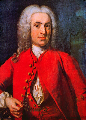
Portrait of Carl Linnaeus in 1739. Author unknown.
Linnaeus returned to Sweden in 1738 and, in Falun, engaged Sara Elisabeth Moraea. They married in 1739 and in 1741 their first son, Carl, was born, and two years later a daughter, Elisabeth Christina, both becoming botanists as their father. The couple had several other children.
During the following years, Linnaeus carried out several expeditions through Europe and, in 1750, became rector of Uppsala University. In 1751, he published Philosophia Botanica, a book containing a survey of his botanical system and other important information on how to do botanical work. In 1753, he published another major work, Species Plantarum, which became internationally known as the starting point of the modern botanical nomenclature. The same year, the King dubbed him knight of the Order of the Polar Star.
His work Systema Naturae continued to be published in updated versions and the 10th edition, published in 1758, became the starting point of modern zoological nomenclature.
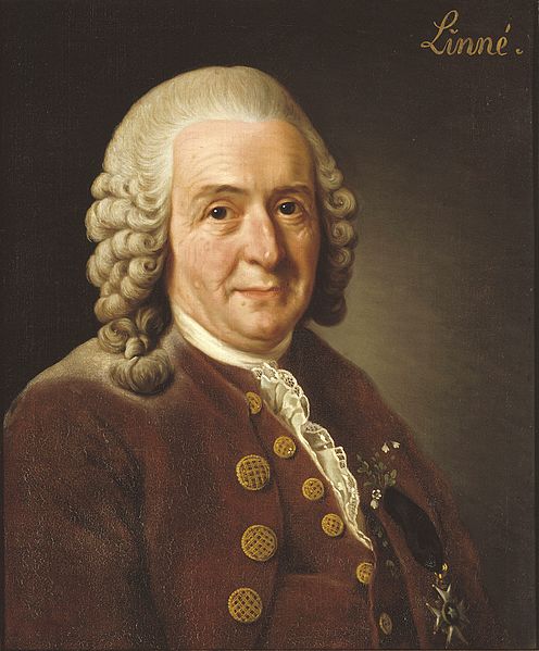
An older Linnaeus in 1775. Portrait by Alexander Roslin.
In 1774, already retired, Linnaeus suffered a stroke that partially paralyzed him. He had a second stroke in 1776, which paralyzed his right side and troubled his memory so that he was unable to identify himself as the author of his own works. A third stroke came in December 1777 and he eventually died on 10 January 1778.
Sorry, guys! It has been about three weeks since my last post, but I was too busy with a lot of personal and academic stuff and wasn’t able to dedicate any time to the blog, but I’m back!
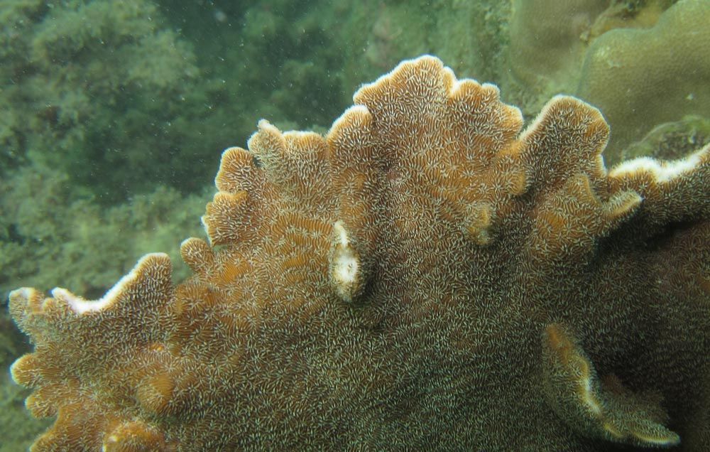
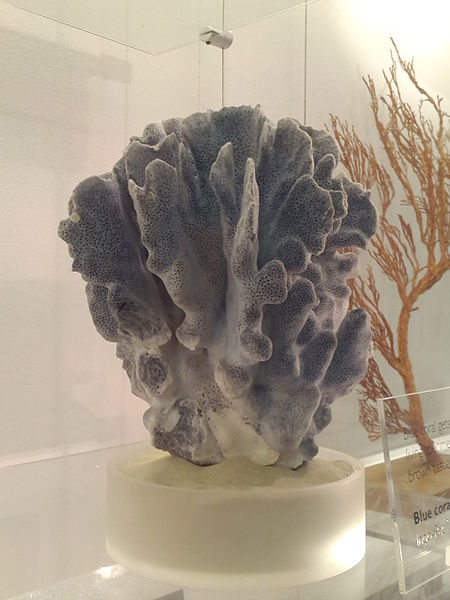
Let’s return with a marine animal as today’s Friday Fellow, the called blue coral, Heliopora coerulea.
A colony of the blue coral in Thailand. Credits to Chaloklum Diving.*
Found in tropical waters of the Pacific and Indian oceans, the blue coral is a peculiar species, being the only one in the genus Heliopora and in the family Helioporidae. It is the only species in the subclass Octocorallia that has a massive skeleton, a feature more common in the stony corals of the subclass Hexacorallia. As a result, the ecological role of the blue coral is usually closer to that of stony corals that to that of its closer relatives.
The skeleton of the blue coral is composed of aragonite and has a distinctive bluish-gray color caused by the presence of iron salts. There are fossils of bluish corals with the same morphology that date back to the Cretaceous, indicating that this is a very old species.
A skeleton of the blue coral in the Natural History Museum, London. Photo by Wikimedia user Kinkreet.**
Although widespread, the blue coral is currently considered a vulnerable species, with some population showing very low genetic diversity. This species is threatened mainly by the jewelry and aquarium trades and by the acidification of the oceans.
Beautiful and deadly, today’s fellow appears during spring as gelatinous orange projections coming out of juniper trees in Europe and North America. Its name is Gymnosporangium sabinae, commonly known as the pear rust.
The jelly-like horns of the pear rust on a juniper tree. Photo by Mark Sadowski.*
The pear rust is a basidiomycete, i.e., a fungus of the phylum Basidiomycota, therefore related to the common mushrooms, but belonging to a different class, the Puccioniomycetes.
During winter, the pear rust remains in a resting state inside branches and twigs of juniper trees. After wet days in spring, the fungus sprouts and appears as horn-like growths covered by an orange gelatinous mass, which are called telia. The telia produce wind borne spores called teliospores that can infect pear trees.
The pear rust on pear leaves. Photo by Jan Homann.
Once reaching the pear tree, the teliospores germinate and infect the leaves of the new host. The infection appears in summer as rust-colored spots on the leaves, hence the name pear rust. In heavily infected plants, the effects of the pear rust can be severe, sometimes causing the plant to lose all its leaves.
In pear trees, the fungus produce reproductive structures known as aecia. They come out from the underside of pear leaves and produce spores called aeciospores, which are able to infect new juniper trees.
The aecia coming out of the rust on a pear tree. Photo by H. Krisp.**
Due to the economic importance of pear trees to humans, the pear rust is a species of great concern. Some countries have policies intended to reduce the spread of the disease, such as preventing transportation of juniper trees from areas known to have the fungus to areas in which it is unknow. In areas where the fungus exist, the solutions to reduce the damage include the use of chemical fungicides, the removal of infected branches in juniper trees and sometimes the removal of any juniper tree around the areas where pear trees are cultivated.
– – –
References:
Fraiture, A.; Vanderweyen, A. (2011) Gymnosporangium sabinae: such a beautifiul disease. Scripta Botanica Belgica11: 193–194.
Ormrod, D. J.; O’Reilly, H. J.; van der Kamp, B. J,; Borno, C. (1984) Epidemiology, cultivar susceptibility, and chemical control of Gymnosporangium fuscum in British Columbia.Canadian Journal of Plant Pahology, 6: 63–70.
Today we celebrate 159 years since the botanist Joseph Maiden was born.
Joseph Henry Maiden was born on April 25, 1859 in London, being the son of Henry Maiden, a china dealer and later accountant, and Mary Elizabeth Maiden (née Wells). In school, he excelled in sciences, later starting to study them in the University of London. However, due to its ill health, he was unable to complete the course.
As part of the treatment of his condition, he was advised to take a long sea voyage, so in 1880 he sailed to New South Wales, Australia, establishing in Sydney, where he at first was invited to deliver a course of lectures by the Working Men’s College. In 1881, Archibald Liversidge (1846–1927), a friend of his chemistry professor, offered him a post of curator at the new Technological Museum in Sydney.
Although intending to return to London within a year, Maiden accepted the job and ended up enjoying it more than he thought he would. This led him to stay longer, and longer, and he started to dedicate himself to the study of the local flora. Unfortunately, in 1882, the Garden Palace, which housed his botanical collection, was destroyed by fire. He, however, started it again with his small staff.
In November 30, 1883, Maiden married Eliza Jane Hammond in Melbourne, which reduced even more his chances of ever going back to England. In 1885, he began to study at the University of Sydney but started to have health issues once more…
Maiden’s interest in Australian flora quickly made him an expert in economic botany. He encouraged the study of the properties of Australian timbers and essential oils. In 1889, he published The Useful Native Plants of Australia. In 1890, he was appointed consulting botanist to the Department of Agriculture and in 1894 became Superintendent of Technical Education.
In 1896, Maiden became Government Botanist and Director of the Botanic Gardens. In this new position, he quickly led to the establishment of Australia’s first herbarium, as well as a museum, a library and a playground.
Joseph Henry Maiden, circa 1900
Maiden became an expert in the genera Acacia and Eucalyptus and his work A Critical Revision of the Genus Eucalyptus, in 8 volumes, remained a major reference for more than half a century. He was also worried about the environment, promoting the preservation of large areas of native forests, and published important works on the use of plants to establish soils and mitigate floods.
In 1924, Maiden retired and moved to Turramurra. He died on November 16, 1925, aged 66, of a heart disease, leaving his wife and four daughters. His only son was lost at sea many years earlier.
– – –
References:
Mark Lyons and C. J. Pettigrew, ‘Maiden, Joseph Henry (1859–1925)’, Australian Dictionary of Biography, National Centre of Biography, Australian National University, http://adb.anu.edu.au/biography/maiden-joseph-henry-7463/text12999, published first in hardcopy 1986, accessed online 25 April 2018.
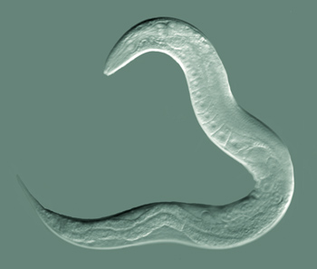
Despite its small size, today’s fellow is one of the most important organisms in current scientific research. Named Caenorhabditis elegans and usually called simply C. elegans, this worm is a nematode and reaches about 1 mm in length and lives in the soil of temperate areas.
An adult hermaphrodite of C. elegans. Photo by Bob Goldstein.*
There are only four bands of muscles that run along the body of C. elegans and they only alow the worm to bend the body dorsally or ventrally, but not to the sides. Thus, while moving on a horizontal surface, the worms are forced to lie on their left or right side.
The main food source of C. elegans are bacteria that live on decaying organic matter, although they can also feed on some yeast species. Therefore, they thrive in soils rich in organic matter, where bacteria occur in abundance.
The sex of C. elegans is unusual. An adult organism can be either a male or a hermaphrodite, without a pure female form. Hermaphrodites are the most common form and usually self-fertilize, although they can, and apparently prefer, to mate with males. The larvae pass through four larval stages before reaching the adult stage, but this happens very quickly, since in ideal conditions the lifespan of C. elegans is of about 2 to 3 weeks. However, in conditions of insufficient food, an alternative third larval stage called dauer can be formed. The dauer stage has the body sealed, including the mouth, which doesn’t allow it to take in food, and can remain as such for a few months until the conditions are good again.
As most nematodes, C. elegans presents eutely, i.e., the adult worm has a genetically determined number of cells in the body. This number is fixed and does not change, because cell division ceases in adults. Male C. elegans have 1031 cells and hermaphrodites have 959 cells.
Due to its small size, small and fixed number of cells, transparent body and because it is easy to raise it in the lab, C. elegans became a perfect model organism. It was the first organism to have its genome fully sequenced and up to now it is the only organism with a complete connectome (the map of his neuron connections). It has been used in studies related to ageing, development, apoptosis and all sort of gene expressions.
Klass, M. R. (1977) Aging in the nematode Caenorhabditis elegans: Major biological and environmental factors influencing life span. Mechanisms of Ageing and Development 6: 413–429. https://doi.org/10.1016/0047-6374(77)90043-4
Peden, E.; Killian, D. J.; Xue, D. (2008) Cell death specification in C. elegans. Cell Cycle 7(16): 2479–2484. https://doi.org/10.4161/cc.7.16.6479
Today is the 187th birthday of the German zoologist Carl Eduard von Martens.
Born in Stuttgard on April 18, 1831, von Martens had three older sisters. His father was a councillor in the Würtemburg Civil Service, but is better known as an explorer of the Fauna and Flora of southern Germany. As both his father and his sisters showed great interest in the natural world, von Martens followed their paths and since an early age he became interested in collecting and studying shells. In school, he was always an astonishing student and showed little interest in playing with the other boys, preferring to dedicate his time to his passion for shells.
Later, he went to Tübingen and studied medicine at the local university. Under the tuition of Hugo von Mohl (1805–1872) and Friedrich August von Quenstedt (1809–1889), von Martens expanded his studies on natural history and became particularly interested in the structure and distribution of animals in space and time. After graduating in 1855, he moved to Berlin, attracted by the fame of Johannes Peter Müller (1801–1858). After returning from a vacation tour to Norway with Müller, von Martens obtained an appointment at the Berlin Zoological Museum (currently the Natural History Museum, Berlin), which at the time was under the direction of Hinrich Lichtenstein (1780–1857). From 1859 on, von Martens was attached to this institution, being assigned to the division of invertebrates (excluding insects). Under his care, the collection of mollusks grew hugely, becoming one of the largest in the world up to the present.
In 1860, von Martens embarked on the Thetis expedition of the Prussian government to Eastern Asia, remaining with the expedition until 1862. When the ship returned to Europe, he remained for two additional years in Asia and studied intesively the local Fauna, especially the mollusks of the Sunda and Moluccas Islands.
Carl Eduard von Martens, around 1901.
During his career, von Martens described almost 1800 animal species, of which 1680 were mollusks, becoming one of the most influential names in malacology.
He died on August 14, 1904, aged 73, leaving a wife and a daughter, as well as an immeasurable contribution to biology.