Friday Fellow: Common Water Bear

by Piter Kehoma Boll

Tiny and tough, our newest Friday Fellow can be found hidden among the moss throughout most of the planet, and perhaps even beyond it, for if there is a species to which space is a piece of cake, that species is the common water bear Milnesium tardigradum.

783px-sem_image_of_milnesium_tardigradum_in_active_state_-_journal-pone-0045682-g001-2

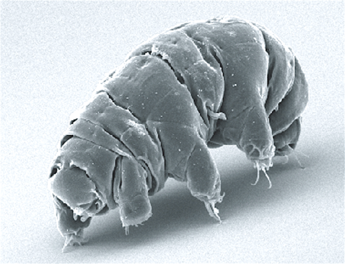
A Scaning Electron Microscope (SEM) image of a specimen of the common water bear in its active state. Photo extracted from Schokraie et al. (2012).*

You may have already heard of tardigrades or water bears, tiny chubby animals that are able to withstand the harsher conditions, such as intense desiccation, radiation and even the vacuum of outer space. Most of the data regarding the toughness of these organisms comes from the common water bear, the most widespread species of the phylum Tardigrada.

Measuring up to 0.7 mm in length, the common water bear has eight legs with claws on their end and is considered a predator, feeding on a variety of other small organisms, including algae, rotifers and nematodes. It has a worldwide distribution and is commonly found living on moss, such as the cosmopolitan silvergreen moss already presented here.

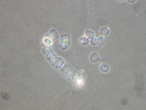
As members of the supergroup Ecdysozoa (which also includes arthropods and roundworms), tardigrades undergo ecdysis, also commonly known as molting, a process through which they shed their exoskeleton. In the common water bear, females always lay eggs around the time of molting. Before leaving the old exoskeleton, the females lay the clutch of eggs, which may vary from 1 to 12 eggs, between the old and the new exoskeleton and usually remain inside the old exoskeleton several hours after laying the eggs. When they finally leave, the eggs remain inside the shed skin, which perhaps helps them to be more protected from danger.

Milnesium_tardigradum

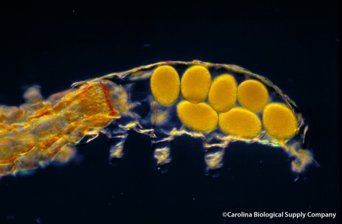
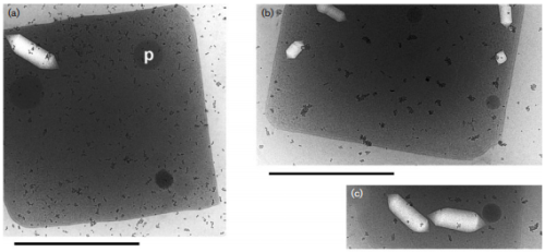
A clutch of seven eggs is left in the empty exoskeleton while the female leaves. Photo by Carolina Biological Supply Company.**

When the habitat of the common water bear gets dry, it enters in a state called cryptobiosis, in which the body shrinks and the metabolism stops. Under this state, known as tun, it can withstand high doses of radiation and both high and zero air pressure, surviving even in the environment of outer space. It is not invincible, however. Radiation in doses above 1000 Gy may not always kill them, but always let them sterile, which is, evolutionary, basically the same thing.

615px-sem_image_of_milnesium_tardigradum_in_tun_state_-_journal-pone-0045682-g001-3

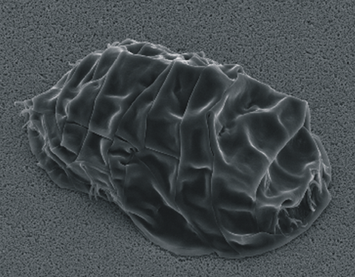
SEM image of the common water bear in the tun state. Photo extracted from Schokraie et al. (2012).*

Nevertheless, the American cockroach is just an amateur regarding survival when compared to the common water bear.

– – –

References:

Horikawa, D. D.; Sakashita, T.; Katagiri, C.; et al. (2009) Radiation tolerance in the tardigrade Milnesium tardigradumInternatonal Journal of Radiation Biology, 86(12): 843–848. http://dx.doi.org/10.1080/09553000600972956

Schokraie E, Warnken U, Hotz-Wagenblatt A, Grohme MA, Hengherr S, et al. (2012) Comparative proteome analysis of Milnesium tardigradum in early embryonic state versus adults in active and anhydrobiotic state. PLoS ONE 7(9): e45682. https://dx.doi.org/10.1371/journal.pone.0045682

Suzuki, A. C. (2003) Life history of Milnesium tardigradum Doyère (Tardigrada) under a rearing environment. Zoological Science 20(1): 49–57. https://doi.org/10.2108/zsj.20.49

– – –

*Creative Commons License
This work is licensed under a Creative Commons Attribution 2.5 Generic License.

**Creative Commons License
This work is licensed under a Creative Commons Attribution-NonCommerical-NoDerivs 2.0 Generic License.

Leave a comment

Filed under Evolution, Zoology

They only care if you are cute: how charisma harms biodiversity

by Piter Kehoma Boll

Which of the two species shown below is more charismatic?

Tangara_chilensis

Tangara chilensis (Paradise Tanager). Photo by flickr user ucumari.*

854px-apocrypta_guineensis2c_volw-wyfie_op_f_sur2c_manie_vd_schijff_bt2c_f

Apocrypta guineensis (a fig wasp). Photo by Wikimedia user JMK.**

You probably would pick the first one. And if I’d ask you which one deserves more attention and efforts to be preserved, you would likely choose the bird as well, or at least most people would. But what is the problem with that? That’s what I am going to show you now.

As we all know, the protection of biological diversity is an important subject in the current world. Fortunately, there is an increase in campaigns promoting the preservation of biodiversity, but unfortunately they are almost always directed to a small subset of species. You may find organizations seeking to protect sea turtles, tigers, eagles or giant pandas, but can you think of anyone wanting to protect beetles? Most preservation programs target large and charismatic creatures, such as mammals, birds and flowering plants, while smaller and not-so-cute organisms remain neglected. And this is not only true in environments that included non-biologist people, but in all fields of research. And more than only leading to a bias in the protection of ecosystems, this preference leads to thousands of understudied species that could bring biotechnological revolutions to humandkind.

In an interesting study published this week in Nature’s Scientific Reports (see reference below), Troudet et al. analyzed the taxonomic bias in biodiversity data by comparing the occurrence of data on several taxonomic groups to those groups’ diversity. The conclusions are astonishing, although not that much surprising. The most charismatic groups, such as birds, are, one could say, overstudied, with an excess of records, while other, such as insects, are highly understudied. While birds have about 200 million occurences above the ideal record, insects have about 200 million below the ideal number. And the situation does not seem to have improved very much along the years.

41598_2017_9084_fig1_html

The bias in interest is clear. The vertical line indicates the “ideal” number of occurrences of each group. A green bar indicates an excess of occurrences, while a red bar indicates a lack of occurrences. Birds and Insects are on the opposite extremes, but certainly the insect bias is much worse. Figure extracted from Troudet et al. (2017).***

Aditionally, the study concluded that the main reason for such disparity is simply societal preference, i.e., the most studied groups are the most loved ones by people in general. The issue is really a simple matter of charisma and has little to do with scientific or viability reasons.

The only way to change this scenario is if we find a way to raise awareness and interest of the general public on the less charismatic groups. We must make them interesting to the lay audience in order to receive their support and increase the number of future biologists that will choose to work with these neglected but very important creatures.

– – –

See also:

Once found and then forgotten: the not so bright side of taxonomy

The lack of taxonomists and its consequences on ecology

Unknown whereabouts: the lack of biogeographic references of species

– – –

Reference:

Troudet, J.; Grandcolas, P.; Blin, A,; Vignes-Lebbe, R.; Legendre, F. (2017) Taxonomic bias in biodiversity data and societal preferences. Scientific Report 7: 9132. https://dx.doi.org/10.1038/s41598-017-09084-6

– – –

*Creative Commons License
This work is licensed under a Creative Commons Attribution-NonCommerical-NoDerivs 2.0 Generic License.

**Creative Commons License
This work is licensed under a Creative Commons Attribution-ShareAlike 3.0 Unported License.

***Creative Commons License
This work is licensed under a Creative Commons Attribution 4.0 International License.

1 Comment

Filed under Botany, Conservation, Systematics, taxonomy, Zoology

Friday Fellow: Pink Cellular Slime Mold

by Piter Kehoma Boll

Protists have always been problematic organisms, and today’s Friday Fellow is not different. In fact, it has probably been one of the most problematic ones. Known scientifically as Acrasis rosea, it has no common name, as you may have guessed already, so I will call him the pink cellular slime mold, as I saw him being called once.

acrasis_rosea_31330

Isolated (or not so much) cells of Acrasis rosea. Photo by Shirley Chio.*

The pink cellular slime mold is a single-celled organism with an amoeboid shape. It feeds on a variety of bacteria and yeasts and is commonly found in decaying plant matter. When the food supply is completely consumed and the cells start to starve, they gather and form a colony that act as a single organism that moves like a plasmodium similar to that of slime molds. For this reason they were originally called cellular slime molds and considered related to other organisms showing a similar behavior, such as those of the genus Dictyostelium.

This plasmodium moves through the formation of “pseudopods”. Eventually the cells start to form a pile reaching up into the air that produce fruiting bodies in the form of branched chains of spores. There is a slight division of labor between stalk and spore, but both groups of cells are viable to produce a new generation.

799px-acrasis_rosea_31095

The chains of spores are visible in this image of the pink cellular slime mold during its plasmodium phase. Photo by Shirley Chio.*

The whole process is similar to what is seen in species of Dictyostelium, but the division of labor and the morphology of the plasmodium and the fruiting bodies are a bit more complex. However, with the advancement of molecular phylogenetics, all the slime mold and cellular slime mold classification fell apart.

While Acrasis was revealed to be an excavate, being closely related to organisms such as the euglenas and parasitic flagellates, Dictyostelium is closely related to the true slime molds, such as the already presented here many-headed slime.

But the excavates are still a problematic group among the protists, and so the real position of the pink cellular slime mold may not be settled yet.

– – –

References:

Bonner, J. T. (2003) Evolution of development in the cellular slime molds. Evolution and Development 5(3): 305–313. http://dx.doi.org/10.1046/j.1525-142X.2003.03037.x

Olive, L. S.; Dutta, S. K.; Stoianovitch, C. (1961) Variation in the cellular slime mold Acrasis rosea*. Journal of Protozoology 8(4): 467–472. https://dx.doi.org/10.1111/j.1550-7408.1961.tb01243.x

Page, F. C. (1978) Acrasis rosea and the possible relationship between Acrasida and Schizopyrenida. Archiv für Protistenkunde 120(1–2): 169–181. https://doi.org/10.1016/S0003-9365(78)80020-7

Weitzman, I. (1962) Studies on the nutrition of Acrasis roseaMycologia 54(1): 113–115.

– – –

*Creative Commons License
This work is licensed under a Creative Commons Attribution-ShareAlike 3.0 Unported License.

Leave a comment

Filed under Friday Fellow, protists

Friday Fellow: Operculate Acrochaete

by Piter Kehoma Boll

Last week I introduced a red alga, the Irish moss. Today I’m bringing another alga, this time a green one, but this is not an ordinary green alga, but a parasite of the Irish moss! So let’s talk about Acrochaete operculata, or the operculate acrochaete as I decided to call it in English, since obviously there would be no common name for an alga parasite of another alga.

Discovered and named in 1988, the operculate acrochaete is an exclusive parasite of Chondrus crispus. The infection occurs when flagellate zoospores of the parasite settle on the outer cell wall of the Irish Moss, where they start their development and digest the cell wall, penetrating the tissues of the host. In sporophytes of the Irish moss, the operculate acrochaete digests the intercellular matrix and spreads through the frond, while in gametophytes the infections remains localized, forming papules. The damages caused by the green alga lead to secondary infections by other organisms, especially bacteria, and the infected fronds end up falling apart, completely degradated.

ccrispus

A frond of the host (Chondrus crispus) to the left and the parasitic Acrochaete operculata that infects its tissues to the right. Photo extracted from chemgeo.uni-jena.de

As mentioned last week, the sporophytes and gametophytes of the Irish Moss have different forms of the polysaccharide carrageenan and this seems to be the reason why the parasite infects both forms differently. The sporophytes have lambda-carrageenan, which seems to increase the virulence of the parasite, while the kappa-carrageenan of the gametophyte seems to limit the green alga’s spread.

Since its discovery, the operculate acrochaete and its interaction with the Irish moss has been studied as a way to both reduce its damage on cultivated crops of the red alga and as a model to understand the relationship of plants and their pathogens.

– – –

References:

Bouarab, K.; Potin, P.; Weinberger, F.; Correa, J.; Kloareg, B. (2001) The Chondrus crispus-Acrochaete operculata host-pathogen association, a novel model in glycobiology and applied phycopathology. Journal of Applied Phycology 13(2): 185-193.

Correa, J. A.; McLachlan, J. L. (1993) Endophytic algae of Chondrus crispus (Rhodophyta). V. Fine structure of the infection by Acrochaete operculata (Chlorophyta). European Journal of Phycology 29(1): 33–47. http://dx.doi.org/10.1080/09670269400650461

Correa, J. A.; Nielsen, R.; Grund, D. W. (1988) Endophytic algae of Chondrus crispus (Rhodophyta). II. Acrochaete heteroclada sp. nov., A. operculata sp. nov., and Phaeophila dendroides (Chlorophyta). Journal of Phycology 24: 528–539. http://dx.doi.org/10.1111/j.1529-8817.1988.tb04258.x


Leave a comment

Filed under Algae, Botany, Disease, Friday Fellow, Parasites

Half male, half female: the amazing gynandromorph animals

by Piter Kehoma Boll

In dioic species, i.e., those in which males and females are separate organisms, sexual dimorphism is very common. It is usually possible to say whether an individual is male or female through external caracteristics, such as color pattern, size or proportion of different body parts.

800px-male_and_female_superb_fairy_wren

Male (left) and female (right) of Malurus cyaneus, the superb fairy-wren. A case of striking sexual dimorphism. Photo by Wikimedia user Benjamint444.*

Vertebrates and arthropods are certainly the two phyla in which sexual dimorphism is best known and found very often. See, for example, the birds above and the spiders below.

521px-male_and_female_a-_appensa

A female (left) and a male (right) of the spider Argiope apensa. The difference in size is more than evident. Photo by Wikimedia user Sanba38.*

The mechanisms that lead to sexual dimorphism are usually the same that lead to the differences in sex by itself. In mammals, birds and arthropods, it is usually due to differences in chromosomes. In other groups, such as crocodiles and snakes, it may be simply a matter of incubation temperature. It is not uncommon to find deviations from this “ideal” dichotomy, with organisms showing unusual chromosome combinations or other features that originate intermediate forms, such as hermaphrodites or androgynous individuals. We have a lot of this in our own species!

There is, however, a much more intriguing and astonishing male-female blend that is often found in arthropods. Known as gynandromorphism, this phenomenon creates specimens with mixed male and female characters forming a mosaic in which one part of the body is male and the other is female. And this distribution is usually bilateral, with one side of the body being male and the other being female.

800px-zwitterhauhechelblaeuling

Gynandromorph of the common blue (Polyommatus icarus). Male on the left side and female on the right side. Photo by Burkhard Hinnersmann.*

800px-heteropteryx_dilatata_0034b_l-d

Gynandromorph of the Malaysian stick insect (Heteropteryx dilatata). Male on the left side and female on the right side. Photo by Wikimedia user Acrocynus.*

A recent paper by Labora & Pérez-Miles (2017) describes the first report of gynandromorphism in a mygalomorph spider (i.e., a tarantula). As the images are not distributed in an open access or creative commons licese, I cannot publish them here, but you can read the article for free thanks to our most beloved god, SciHub.

The causes of gynandromorphism are not always clear, but most of the times it seems to be due to a chromosome impairment in mitosis during the first stages of embryonic development. Thus, it is more likely to occur in inviduals that were originally heterogametic, i.e., they had two different sex chromosomes in their zygote.

Gynandromorph

A gynandromorph cardinal (Cardinalis cardinalis). Photo by Gary Storts.**

Gynandromorphism should not be confused with chimerism, a somewhat similar phenomenon in which an individual is the result of the fusion of two different embryos.

Now tell me, isn’t nature fascinating in every single detail?

– – –

References and further reading:

Jones, S. R.; Philips Jr., S. A. (1985) Gynandromorphism in the ant Pheidole dentata Mayr (Hymenoptera: Formicidae). Proceedings of the Entomological Society of Washington, 87(3): 583–586.

Laborda, A.; Pérez-Miles, F. (2017) The first case of gynandry in Mygalomorphae: Pterinochilus murinus, morphology and comments on sexual behavior.  Journal of Arachnology, 45(2): 235–237. https://doi.org/10.1636/JoA-S-049.1

Labruna, M. B.; Homem, V. S. F.; Heinemman, M. B.; Ferreira Neto, J. S. (2000) A case of gynandromorphism in Amblyomma oblongoguttatum (Acari: Ixodidae). Journal of Medical Entomology, 37(5): 777–779.

Olmstead, A. W.; LeBlanc, G. A. (2007) The environmental-endocrine basis of gynandromorphism (intersex) in a crustacean. International Journal of Biological Sciences 3(2): 77–84.

– – –

*Creative Commons License
This work is licensed under a Creative Commons Attribution-ShareAlike 3.0 Unported License.

**Creative Commons License
This work is licensed under a Creative Commons Attribution-NoDerivs 2.0 Generic License.

Leave a comment

Filed under Entomology, Spiders

Friday Fellow: Irish Moss

by Piter Kehoma Boll

Growing abundantly along the North Atlantic coasts, our newest Friday Fellow is a cartilaginous red alga commonly known as Irish moss or scientifically as Chondrus crispus, which means something like “curly cartilage”.

chondrus_crispus

The Irish moss usually appears as a mass of curly cartilaginous and soft seaweed with a red or purple tinge. Photo by Wikimedia user Kontos.*

Reaching about 20 cm in length, the Irish moss is attached to the substrate by a discoid base and its thallus branches dichotomously four or five times. The width of the branches may vary from about 2 to 15 mm and the color is even more variable, ranging from green or yellowish to dark red, purple, brown or even white. As with all plants, the Irish moss has a gametophyte (haploid) and a sporophyte (diploid) form. The gametophytes have a blue iridescence (as seen in the photo above), while the sporophytes show a dotted pattern (seen above as well).

The Irish moss is edible and relatively well known among the communities living where it grows. In Ireland and Scotland, it is boiled in milk and sweetened to produce a jelly-like product. The cartilaginous or jelly-like appearance of this alga and its derivatives are due to the presence of high amounts of carrageenan, a polysaccharide that is widely used in food industry as a thickening and stabilizing agent and as a vegan alternative to gelatin.

Due to its economic importance, the Irish moss is cultivated in tanks for the extraction of carrageenan and other products. Both gametophytes and sporophytes produce carrageenans of different types that can be used for different purposes.

– – –

References:

Chen, L. C.-M.; McLachlan, J. (1972) The life history of Chondrus crispus in culture. Canadian Journal of Botany 50(5): 1055–1060. http://doi.org/10.1139/b72-129

McCandless, E. L.; Craigie, J. S.; Walter, J. A. (1973) Carrageenans in the gametophytic and sporophytic stages of Chondrus crispus. Planta 112(3): 201–212.

Wikipedia. Chondrus crispus. Available at < https://en.wikipedia.org/wiki/Chondrus_crispus >. Access on August 1, 2017.

– – –

*Creative Commons License
This work is licensed under a Creative Commons Attribution-ShareAlike 3.0 Unported License.

1 Comment

Filed under Algae, Botany, Friday Fellow

Friday Fellow: Baker’s Yeast

by Piter Kehoma Boll

Living along humans for centuries, today’s Friday Fellow is certainly one of the most beloved fungi. Scientifically known as Saccharomyces cerevisiae, its common names in English include baker’s yeast, brewer’s yeast or ale’s yeast.

800px-saccharomyces_cerevisiae_sem

Saccharomyces cerevisiae under the scanning electron microscope. Photo by Mogana Das Murtey and Patchamuthu Ramasamy.*

Under the microscope, the cells of this single-celled species are ellipsoid or sphere-shaped and usually show small buds from new cells growing from the larger one. But you may have seen this species being sold as tablets or grains in the supermarket, as they are used to make bread and many alcoholic bevarages, such as wine and beer, but the baker’s yeast is much more interesting than just that.

450px-d094d180d0bed0b6d0b6d0b8_d181d183d185d0b8d0b5_d0b1d18bd181d182d180d0bed0b4d0b5d0b9d181d182d0b2d183d18ed189d0b8d0b5_-_rapid-rise_yeast

Grains of dried but yet alive baker’s yeast as it is sold commercially.

The cells of the baker’s yeast occur naturally on ripe fruits, such as grapes, and this was likely the original source of the strains currently cultivated by humans. The yeast reaches the fruits through many wasp species that have it growing in their intestines, an ideal environment for the fungus’ sexual reproduction.

As it is easily cultivated in the lab and has a short generation time, the baker’s yeast has become one of the most important model organisms in current biological studies. It was, in fact, the first eukaryotic organism to have its whole genome sequenced more than 20 years ago.

Saccharomyces_cerevisiae

Saccharomyces cerevisiae growing on solid agar in the lab. Photo by Conor Lawless.**

More than giving us food and drink, this amazing yeast has increased our understanding of gene expression, DNA repair and aging, among many other things. Live long the yeast!

– – –

References:

Giaever, G.; Chu, A. M.; Ni, L.; Connelly, C. et al. (2002) Functional profiling of the Saccharomyces cerevisiae genome. Nature 418 (6896): 387-391.

Herskowitz, I. (1988) Life cycle of the budding yeast Saccharomyces cerevisiae. Microbiological Reviews 52 (4): 536-553.

Wikipedia. Saccharomyces cerevisiae. Available at < https://en.wikipedia.org/wiki/Saccharomyces_cerevisiae >. Access on July 25, 2017.

– – –

*Creative Commons License
This work is licensed under a Creative Commons Attribution-ShareAlike 3.0 Unported License.
**Creative Commons License
This work is licensed under a Creative Commons Attribution 2.0 Generic License.

1 Comment

Filed under Friday Fellow, Fungi

Old Italian trees: a step toward worldwide recognition?

by Piter Kehoma Boll

Some years ago I wrote an article (you can read it here) about the importance of trees, especially old trees, and how their ecological role is different from that of a young tree.

old_tree

Ancient trees are ecological preciosities and need to be preserved for the sake of their ecosystems. Photo by flickr user loshak.*

In Italy, there are specific laws erected to protect ancient trees, especially if they are unique for their species or have some sort of aesthetic or cultural value. Recently, their importance for the preservation of a variety of lifeforms has also started to be recognized. A recently published article by a group of Italian researchers (see below) compared the noteworthy old trees in Italy recorded in a previous list and a new list. They conclude that the new inventory has considerable improvements, although some issues remain, including the presence of exotic, even invasive, species in the list.

But such initiatives are at least important as a first step that may guide us to a better understanding and management of old trees, which are precious elements, but continue to decline worldwide.

Read the study for free:

Zapponi, L.; Mazza, G.; Farina, A.; Fedrigoli, L.; Mazzocchi, F.; Roversi, P. F.; Peverieri, G. S.; Mason, F. (2017) The role of monumental trees for the preservation of saproxylic biodiversity: re-thinking their management in cultural landscapes. Nature Conservation 19: 231–243.

– – –

*Creative Commons License
This work is licensed under a Creative Commons Attribution-NonCommerical-NoDerivs 2.0 Generic License.

Leave a comment

Filed under Conservation, Ecology

Friday Fellow: Walsby’s Square Haloarchaeon

by Piter Kehoma Boll

After more than a hundred Friday Fellows, there is still one group with no representatives here: the archaeans. But this is going to change today with the indroduction of our first Friday Fellow archaean, and it is a very interesting one for sure.

Scientifically known as Haloquadratum walsbyi, it is sometimes called Walsby’s Square Haloarchaeon and, as its name suggest, it has an unusual square shape.

haloquadratum_walsbyi00

A drawing showing a set of four cells of Walby’s square haloarchaeon.

This interesting archaean was discovered in 1980 by Anthony Edward Walsby in brine ponds of the Sinai Peninsula. It was later discovered in several other lakes with high concentrations of salt around the world and was first cultivated in the laboratory in 2004, but only in 2007 it was formally described and received a binomial name.

The square cells of the Walsby’s square haloarchaeon are very thin, about 0.2 µm thick, and measure about 2 µm on each side. They grow very slowly, forming a thin sheet over a surface, the largest recorded sheet measuring 40 × 40 µm. If the growing conditions are not ideal, the cells deteriorate to a ragged square or other shapeless flat form.

Haloquadratum

Photographs of cells of Haloquadrum walsbyi showing the crystal-shaped air vacuoles. Image extracted from Burns et al. (2007).

Inside the cells, the Walsby’s square haloarchaeon has small gas vesicles that look like small crystals. They help the cell remain at the surface of the very salty water they inhabit. In order to survive, this archaean needs water with a concentration of salt of at least 14%, but the conditions become ideal only above 23%.

Although we know some interesting things about this species, there is still much more to learn. Who knows what mysteries this small square-shaped creature is hiding from us?

– – –

References:

Bolhuis, H.; Poele, E. M. t.; Rodriguez-Valera, F. (2004) Isolation and cultivation of Walsby’s square archaeonEnvironmental Microbiology 6(12): 1287–1291.

Burns, D. G.; Janssen, P. H.; Itoh, T.; Kamekura, M.; Li, Z.; Jensen, G.; Rodríguez-Valera, F.; Bolhuis, H.; Dyall-Smith, M. L. (2007) Haloquadratum walsbyi gen. nov., sp. nov., the square haloarchaeon of Walsby, isolated from saltern crystallizers in Australia and SpainInternational Journal of Systematic and Evolutionary Microbiology 57: 387–392.


Leave a comment

Filed under Archaeans, Friday Fellow

Friday Fellow: Tiger Worm

by Piter Kehoma Boll

European in origin, but currently cosmopolitan, today’s Friday Fellow is a very useful earthworm for humans. Scientifically known as Eisenia fetida, this species has many different popular names, including tiger worm, red californian earthworm, red wiggler worm, etc.

Eisenia_fetida

Two specimens of Eisenia fetida. Photo by iNaturalist.org user nzwormdoctor.*

The tiger worm rarely lives underground, prefering to live among decaying vegetable matter, such as in the leaf litter, therefore being considered an epigean species. Due to its adaptability to live among and feed on decaying organic material, it is widely used by humans for vermicomposting, i.e., producing humus to be used as a nutrient rich soil in cultivation of vegetables. As a result, it has been introduced worlwide.

When molested, the tiger worm secrets a yellow and pungent liquid from its celomic cavity that has been shown to be toxic to some vertebrates, thus probably being a defense mechanism against predators.

Due to its agriculatural importance, the tiger worms has been used in many studies regarding its response to different soil contaminants, including pesticides, and its presence on the amount of inorganic nutrients, such as carbon and nitrogen, in the soil.

– – –

References:

Albanell, E.; Plaixats, J.; Cabrero, T. (1988) Chemical changes during vermicomposting (Eisenia fetida) of sheep manure mixed with cotton industrial wastes. Biology and Fertility of Soils, 6(3): 266–269.

Spurgeon, D. J.; Hopkin, S. P. (1999) Comparisons of metal accumulation and excretion kinetics in earthworms (Eisenia fetida) exposed to contaminated field and laboratory soils. Applied Soil Ecology, 11(2–3): 227–243.

Zhang, B.-G.; Li, G.-T.; Shen, T.-S.; Wang, J.-K.; Sun, Z. (2000) Changes in microbial biomass C, N, and P and enzyme activities in soil incubated with the earthworms Metaphire guillelmi or Eisenia fetidaSoil Biology and Biochemistry, 32(14): 2055–2062.

– – –

*Creative Commons License
This work is licensed under a Creative Commons Attribution-NonCommercial-ShareAlike 4.0 International License.

Leave a comment

Filed under Friday Fellow, worms